The University of Southampton

Unpacking the Nuremberg Code’s Legacy in Medical Research

Since 1947, a set of ten standards has existed to prevent harm to human subjects during clinical trials, following the infamous Nazi human experiments that took place during World War II in concentration camps. These horrific trials left many dead, and almost all survivors experienced permanent severe injuries. The crimes included transplantation, amputation, starvation, freezing and sterilization amongst others, and any victims who stood against the doctors were sent to the gas chambers. As much as it can be unanimously agreed that this is one of the greatest examples of brutal medical malpractice, it can be surprisingly difficult to unpack the ethics of using the collected knowledge in the 21st century for the advancement of modern health.

Jadwiga Dzido showing her scars from medical experiments

There are two major arguments against using the data obtained from the experiments. Over the years, the data has been used in multiple fields. I would like to focus on the Dachau freezing experiments, which are possibly the most controversial due to their prevalence in 20th-century research publications into the treatment of hypothermia. This involved ‘participants’ being submerged in tanks of ice water, some anesthetised, others conscious, to induce hypothermia, so that rewarming techniques could be tested on their efficacy and bodily responses. I use the term ‘participants’ extremely loosely, since this was done without consent, usually forced or under the false pretence of release from concentration camps.

Argument one considers the actual value of the data collected regardless of its ethics. Physician-scientist Andrew Ivy declared ‘Nazi experiments on humans were of no medical value’ at the Nuremberg war crime trials. However, recently, several investigators have suggested the Dachau study did produce credible data. Understandably, there is no ethical way to generate the data produced from this study, and important understandings on treating hypothermia could be life-saving. This is where argument two gains significance. The data gathered could potentially be considered ethically usable if it is for the greater good.

The experiment should be such as to yield fruitful results for the good of society, unprocurable by other methods or means of study, and not random and unnecessary in nature

– The Nuremberg Code

Conclusion

Now, the Dachau experiments completely go against all but one of the ten postulates of the Nuremberg Code. There was no informed voluntary consent, injury and suffering was certainly not avoided and there was absolutely no opportunity for the human subjects to terminate the trials. In every sense of the word, these experiments were not ethical. Whether they should have occurred is not up for debate. But since replication of these experiments is off the table, and lives could be saved with this knowledge, could it be suggested that the saving of lives is almost … honouring those who died and suffered? Equally, however, it could be argued that continuing to revoke their ability to consent is entirely dishonourable. In my opinion – in an ideal world, these experiments would never have happened. The 15,754 documented victims died needlessly. Despite this, if it can be agreed that the data collected is of scientific relevance and value, could be used for the greater good, and cannot be ethically replicated, this data should not go to waste, along with the legacy of the victims.
Stem cells: Science’s golden egg?

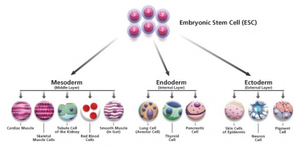
Every cell within our body begins as a stem cell, from immune cells to our neurons and play an integral part of human development. There are many different types of stem cell, which are divided up into 4 classes: totipotent, pluripotent, multipotent and unipotent. Totipotent stem cells are cells that can differentiate into any type of cell, but pluripotent cells can differentiate into most, but not all cell types. Multipotent stem cells are more specific than pluripotent stem cells, but can still differentiate into a few different cell types, and Unipotent stem cells can only become one type of stem cell. Pluripotent stem cells or more specifically embryonic stem cells, are most widely used in stem cell treatment.

A diagram of the cells that an embryonic stem cell can create

So how are these cells currently being clinically used?

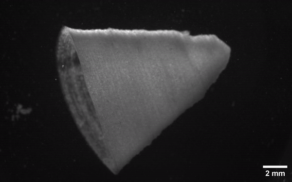
Pluripotent stem cells are currently being used in a host of different ways, with excellent research into the treatment of neurodegenerative diseases like Parkinson’s and many others. Stem cells have also been used to 3D print heart beating cells, as well as entire tissues. These groundbreaking discoveries only begin to scratch the surface of the incredible research that is being put into stem cells.

Picture of the 3D printed beating heart cells

What does the future hold?

Although scientists are currently struggling to use stem cells for more intricate structures, like limbs, which require multiple tissue types in order to function, I believe that in the direction in which stem cell research is going and with the unbelievably rapid rate in which it is progressing, that this will eventually be possible. I also believe that the 3D printing of tissues using stem cells will eventually allow for the 3D printing of entire organs, meaning that organ transplants are no longer needed, also nullifying the extremely long waitlists some people have to endure in order to receive a transplant, or who may not even receive a transplant at all. It would also completely eradicate any immune rejection that would occur in an organ transplant. I think stem cells are truly the ‘miracle cure’ for disease, and would revolutionise clinical treatment as we know it. I am extremely excited to see how this research develops and how much we can achieve. Stem cells really are science’s golden egg.

Are stem cells worth it?

Currently, there is a lot of debate about the ethics behind the use of stem cell therapy, specifically using embryos to harvest them. This has sparked an extremely polarising debate about when embryos are considered morally human, as since some people believe that, from the moment of fertilisation, the embryo is morally human and that destroying the embryo in order to retrieve these stem cells is murder. This very much shows a Kantian view of ethics, in which he describes ethics as what is right and questions the morality of the action. However, Jeremy Bentham’s view of ethics which is that what is good is what has good outcomes, which brings into question whether the action of destroying embryos outweighs the outcome of the production of revolutionary treatments and medical care. I believe that, as the moral status of embryos is still being debated, I don’t think that the progress which is currently being made in stem cell therapy should be halted. However, I do believe that strict regulations in to how long an embryo can be grown should be put into place to minimise exploitation and unethical behaviour by allowing for the embryo to begin developing much further.

The Stem Cell Saviour

Stem cells have always been an intriguing topic to me, particularly their therapeutic potential. The idea of being able to use one undifferentiated cell to create whole new organs or body parts is fascinating. After our lecture on stem cells and their therapeutic use, I decided to research the different ways that stem cells are currently being used in medicine, and one case in particular stood out.

The Story

Lesley Calder was diagnosed with acute myeloid leukaemia in 2019. Chemotherapy treatment was unsuccessful for her cancer, and she was left with the only option of a stem cell transplant. Lesley’s three siblings volunteered to be tested, with the slim chance of finding a sibling match (~25%). By some miracle, 2 siblings were full matches and 1 was a half match. Lesley’s sister Annie was chosen to be the donor, and amazingly, Lesley has since made a full recovery.

Using Stem Cells to Treat Cancer

A stem cell is defined as a cell that can self-renew indefinitely and has the capacity to differentiate into many cell types. Their normal function within the body is to replace old, damaged or defective cells to maintain normal tissue function.

Stem cell transplants are used to treat diseases where the bone marrow is damaged or defective, meaning that healthy blood cells can no longer be produced. This is the case in blood cancers (e.g. leukaemia and lymphoma), which primarily affect white blood cells. The loss of blood cells is further exacerbated by intensive cancer treatment (e.g. chemotherapy), which can also damage/destroy healthy cells. The transplantation of stem cells produces new blood cells, and helps to defend against the cancer.

Dr Sonali Smith, M.D. explains the process of using stem cells to treat cancer.

Stem Cell vs. Bone Marrow Transplants

Before this research, I had only heard about bone marrow transplants, and didn’t know stem cell transplants existed, which made me wonder what the difference is. They are essentially the same thing, but differ in the locations where the cells are collected. A stem cell transplant involves collecting stem cells from the bloodstream, which is less invasive than a bone marrow transplant, which involves collecting a person’s bone marrow from within their bone (usually pelvic).

Information from Cancer Research UK says that stem cell transplants are the more common of the two, which I found surprising, considering I hadn’t heard of them. This is because stem cell transplants are: less invasive, easier to perform, have a higher yield of cells and have a quicker blood count recovery.

Dr Scott Bearman, M.D. explains the difference between stem cell and bone marrow transplants.

The Problem & Final Thoughts

Through this research, I have found that over 70% of patients who require a stem cell transplant will not find a compatible donor in their family. Additionally, only 3% of the UK population (and <6% of people in Northern Ireland) are registered to be stem cell donors, making the chances of finding a compatible match even lower. For the majority of patients, like Lesley, a stem cell transplant is their only chance at recovery, and their chances of success are slim.

Lesley’s story prompted her son Max to join the stem cell donor register in hopes of helping others like his mum, and he has already been called upon to donate. By sharing her story, I hope to inspire others to join the register as well, as I will definitely be doing. Anyone aged 17-55 and in good health can sign up here. For more information on stem cell transplants, visit NHS, Cancer Research UK or Leukaemia & Lymphoma Society.

Breaking Boundaries: Evolution of Skeletal Muscle Tissue Engineering – From Traditional to 3D Printing Innovation

Approximately 45% of the mass of the human adult body is muscle tissue.

Serge Ostrovidov, PhD et al.

Muscles are critical in locomotion, prehension, mastication, ocular movement, and other dynamic activities such as body metabolic regulation. Myopathy, traumatic injury, aggressive malignant tumour extraction, and muscular denervation are the most prevalent clinical indications for therapeutical or cosmetic reconstructive muscle surgery. There are three types of muscle tissue: smooth muscles in the stomach and intestines, cardiac muscle in the heart, and skeletal muscle throughout the body. Skeletal muscle, which we will focus on in this blog, majorly consists of myocytes, the multinucleated contractile muscle fibres.

Methodology of SMTE

While Skeletal muscle tissue engineering (SMTE) has been introduced to the world of biomedical engineering for more than two decades, it remains a significant challenge, with numerous techniques being developed constantly to accomplish this.

The muscle cells are first isolated from the patient or a donor, followed by culturing, where the cells effectively differentiate and develop in the nutrition-rich medium. The muscle tissue is then generated in three methods, which form different specialized muscle cells: myotube formation under controlled topography, cell sheet formation with a hydrogel matrix, and muscle bundle formation. The well-developed and functional tissue is then transplanted back to the patient.

Limitations

This is the traditional way of engineering skeletal muscle tissue, but it still faces some limitations. Though cells could be isolated from the patient, the tissues generated from a donor could trigger immune responses in the recipient’s body, causing rejection and the necessity for immunosuppressive drugs. These medications come with their own risk, like increased susceptibility to infections and organ damage. Moreover, traditional tissue engineering often focuses on preparing damaged muscle tissue rather than promoting regeneration. Thus, the regenerative potential of the SMTE tissue may be insufficient to restore full muscle function, especially in cases of extensive injury or degeneration quantitatively and qualitatively due to ageing. On the other hand, ensuring the long-term functionality of transplanted muscle tissue is also a significant challenge. Without adequate vascularization and support structures, it may encounter degradation or necrosis over time, leading to repeated interventions.

3D Print Muscle Tissue

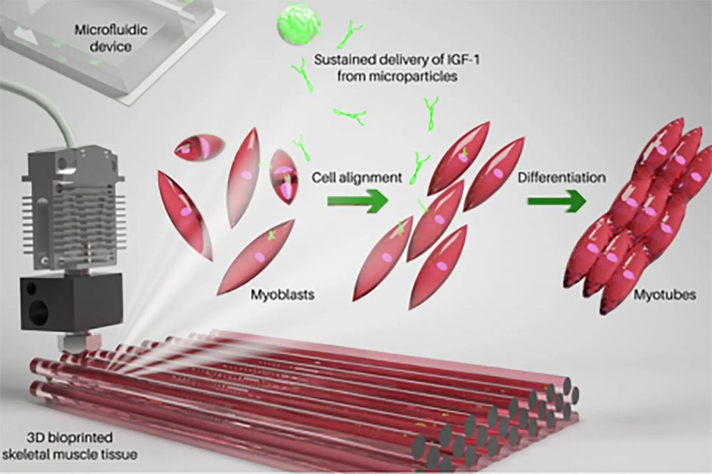
I came across a piece of news published in 2023 written by Michael M., mentioning the researchers at the Terasaki Institute for Biomedical Innovation have developed a fascinating new technology of 3D printing skeletal muscle tissues with bioink invented by the researchers, which could mimic the natural muscle formation.

Photo credits: Terasaki Institute

The science of muscle 3D printing

The three parts of the bioink are polylactic acid (PLGA) microparticles, myoblast cells, and a hydrogel based on gelatin. The PLGA microparticles sustainably release growth factor-1 (IGF-1), an insulin-like hormone that’s necessary for healthy bone and tissue growth. The production process of muscle tissue is more complex than it looks. Myoblast cells were deemed viable three days after printing and successfully developed into complete synthetic muscle tissue over the next ten days, eventually contracting on their own, just like normal muscle tissues! In order to determine if the cultured tissue was viable in living organisms, the tissue was implanted into mice. The researchers were then able to confirm that the cultured tissue was successfully accepted by the body and combined with the existing muscle tissues.

This video shows 3D-printed myotubes, which eventually form muscle fibres, twitching spontaneously after receiving a dosage of the hormone IGF-1.

Summary

The ability to 3D print muscle tissue represents a significant advance in medicine. Using this technology, muscle mass replacement could be revolutionized by mimicking the natural process of muscle development. Prior to the possibility of treating humans, further clinical studies and testing will be necessary to confirm the safety of this process before the possibility of treating humans. What’s for sure, however, is that 3D printing muscle tissue has a highly promising future in medicine. I, as a biomedical science student, am also looking forward to the further development of skeletal muscle engineering.

References:

  1. Ostrovidov, S., PhD et al. (2014). Skeletal Muscle Tissue Engineering: Methods to Form Skeletal Myotubes and Their Applications. Tissue Engineering Part B Review, 20(5), pp. 403-436. doi: 10.1089/ten.teb.2013.0534

DIY Artificial Pancreas: a game changer for diabetic care?

During our sensor lectors, the concept of an artificial pancreas was discussed. This stood out to me as my stepmother has diabetes and I had never really considered how having this actually affected her life. It also made me aware that I didn’t even know if she already used an artificial pancreas! Then through researching, I was drawn to the DIY artificial pancreas. I wanted to understand what this was and whether it was the most beneficial option in terms of cost, function and aesthetics.

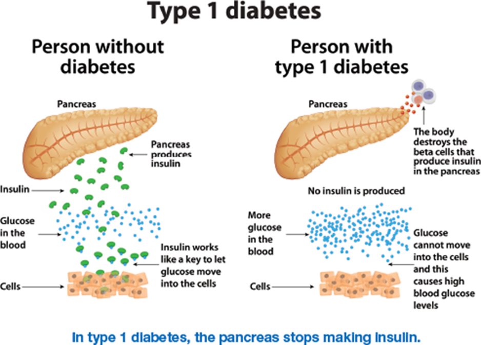
Individuals with Type 1 diabetes cannot produce insulin to monitor their blood glucose levels. Instead, they inject insulin throughout the day; calculating how much is needed dependent on factors such as what they’ve consumed.

Insulin production for a healthy individual vs an individual with Type 1 diabetes https://www.ndss.com.au/about-diabetes/type-1-diabetes/

What is an artificial pancreas and how does it work?

An artificial pancreas (also known as a closed loop system) can monitor blood glucose levels automatically, calculating and administering the required amount of basal (background) insulin. This is comprised of three components: a continuous glucose monitor (CGM) which uses tiny sensors under the skin that track the blood glucose levels every few minutes, delivering information wirelessly to an insulin infusion pump to administer insulin when the blood glucose levels is not within the target range as well as to a program on a device such as a phone to monitor the readings.

This image demonstrates the components
of the artificial pancreas and how they work.
https://www.bbc.co.uk/news/health-60133358

What is a DIY artificial pancreas and how does it differ?

A DIY artificial pancreas is non-NHS funded but uses similar equipment. It uses a specific app to control your equipment which needs to be compatible with your insulin pump. Guidance is mainly provided through the diabetes community and not the NHS as they have limited knowledge on how these programs work. If you are’ techie’ then you can fine tune this program and ‘train’ the system: making this an advancement on the NHS funded system.

What are the pros and cons?

Now, there are benefits to this: insulin is given automatically when needed which can be ideal especially for young children; allowing a more stress-free experience whilst giving their parents a way to monitor their blood glucose levels throughout the day. It also removes the need to inject which can be beneficial for those who dislike needles. As mentioned, the DIY version can be ‘trained’ to your specific needs so will reduce the energy and brainpower the individual spends on calculating the correct levels. However having said that, because it is technology, this can always malfunction and if given the wrong amount of insulin, it can cause a hypo. The program also requires internet connection where you still need to input your meals as the artificial pancreas can only give basal insulin automatically. In terms of the DIY version, it can be complicated to install and needs updating regularly. Since it needs to be attached to either a belt, a belt loop or a pouch, aesthetically the insulin pump can look bulky under clothing and limit the options of available outfits an individual could wear. This could cause insecurities or just be an added inconvenience.

Video discussing my stepmother’s sensors vs a DIY artificial pancreas

After both points of views it’s clear how life changing a DIY artificial pancreas can be!

‘Watch’ this space: how our favourite devices could detect Alzheimer’s disease.

What is Alzheimer’s Disease?

Having close personal experience with dementia, I often think about the subtle changes in behaviour and function that I noticed, but initially overlooked, in family members who were (much) later diagnosed. Alzheimer’s disease (AD) is the most common form of dementia and accounts for 60-80% of dementia cases(1). It is a degenerative brain disease, where cell damage leads to complex brain changes that gradually worsen over time. Crucially, the slow progression of AD means that behavioural and physiological signs, or ‘biomarkers’ may present long before a diagnosis is even considered. I imagine many people who care for or love someone with dementia feel similarly, particularly as AD currently has no effective treatment and the current method of clinical diagnosis is criticised for cultural bias and inaccuracy. Early diagnosis is essential as the chance of reversing anatomic and physiologic changes decreases dramatically as the disease advances(2). Personally, I believe one of the most important benefits of early diagnosis is allowing people with dementia to open a dialogue about their ideal care plan, and express autonomy over their future.

The inspirational Wendy Mitchell

I highly recommend reading “What I wish People knew about dementia” by Wendy Mitchell, which provides a great insight into how early diagnosis and acceptance of dementia can help individuals maintain independence and autonomy.

Towards Digital Detection

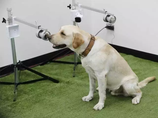
A labrador retriever trained to sniff out covid

The race to improve diagnosis and treatment for AD is on. In 2023 there was even a $200,000 reward advertised for anyone who can prove that dogs can sniff out Alzheimer’s disease. Whilst MRI and PET molecular imaging of beta-amyloid and tau proteins are perhaps more promising, the cost and invasive nature of such methods precludes practical clinical applications(3).

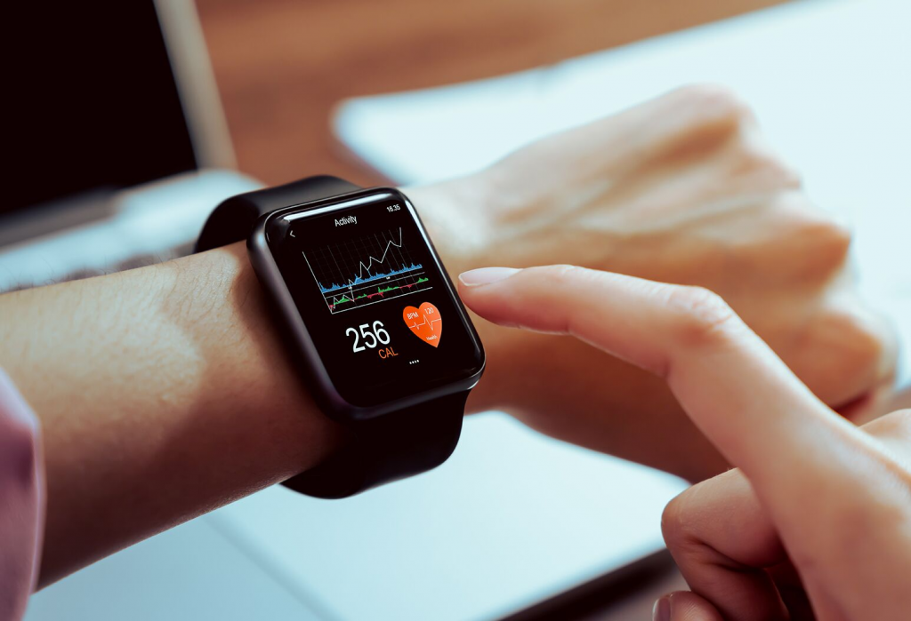
Interestingly, digital consumer technology can overcome these limitations with the added benefit of pre-symptomatic detection. What’s amazing is that cognitive, behavioural, sensory, and motor biomarkers can aid detection of AD 10 to 15 years prior to effective diagnosis(4). This allows leverage of existing technologies and sensors such as smartphone microphones and GPS systems.

Watch this quick video I put together to find out more…

Original video, inspired by Kourtis et al (2019).

Innovation or Invasion?

Whilst I do own both a smartphone and an (albeit old) smartwatch, the concept of these devices continuously monitoring everything from my speech to my walk and even the way I view an Instagram post, makes me slightly uneasy. However, I can’t deny the advantages of personal continuous monitoring for public health. The question is where do we draw the line? Could we be heading down a path where continuous passive monitoring involves cameras wired up in our home, even in our toilets?! (apparently yes! Find out more here).

I think the answer is purely personal – for some people continuous passive monitoring may be the difference between life and death, for others it might feel a little too 1984. For people experiencing cognitive decline, informed consent may complicate matters further. According to the Mental Capacity Act (2005), many people with dementia may be considered ‘Incompetent adults’ (I’m not a fan of that term) if they fail to understand the device, and cannot remember or communicate the reasoning for use. This means they would be legally unable to consent. Thankfully, the increasing nature of wearable technologies may mean that, in the future, many people will own devices decades before they are at high risk of AD, and thus can choose (at legal capacity) whether or not to install bio-monitoring software. Of course, if these devices continue to be solely commercial, then the financial accessibility of these devices may be limited – which is a whole other debate in itself.

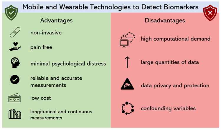
Original image: the pros and cons of digital biomarkers.

Summary

Given the increasingly technological cultural landscape – our access to devices capable of passive monitoring is increasing. Considering the UK Alzheimer’s epidemic and our ageing population, it seems a waste to not make use of the endless potential health benefits of these devices (especially as many already monitor us for consumer metrics anyway)! Although the degree of monitoring may seem invasive, I think Alzheimer’s is a far bigger threat to our personal privacy and autonomy. Such developments could help people communicate with their families and manage symptoms before its too late.

References

  1. Dementia vs. Alzheimer’s Disease: What Is the Difference? | alz.org
  2. Kourtis, L.C. et al. 2019. Nature. 2, 9. https://doi.org/10.1038/s41746-019-0084-2
  3. Bao, W. et al. 2021. Front. Aging Neurosci. https://doi.org/10.3389/fnagi.2021.624330
  4. Vrahatis, A. G. 2023. Sensors. 23, 9. https://doi.org/10.3390/s23094184
  5. Stringer, G. et al. 2018. Int J Geratr Psychiatry. 33,7. https://doi.org/10.1002%2Fgps.4863
  6. Sun, J. et al. 2022. Sec. Brain Imaging and Stimulation. 16. https://doi.org/10.3389/fnhum.2022.972773