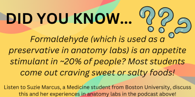
The corridors of the Education wing of the hospital were long and dimly lit, as though the very building bore the solemnity and calm monotony of its workers inside. Apron on and into the lab I went, greeted by the sharp air which washed the senses and clung to clothes; formaldehyde would be the odour to my education for that day. I explored specimens laid out in-front of me through gloved hands, my nerves dissipating in the pursuit of knowledge and yet a silent edge to the room remained, the taboo feeling still choking speech.
My first time stepping into an anatomy lab was an experience like no other. It’s a moment filled with a complex blend of emotions; curiosity, apprehension, and a profound sense of respect for the journey upon which you’re about to embark. Reflecting on this; the presence of the surgeon, his demeanour, his depth of experience and the way he handled the specimens with such delicate precision put me at a surprising sense of ease. It allowed me to see past extraneous detail and appreciate the beauty in the anatomy and the implants used to restore vital function. This kinaesthetic experience cemented the knowledge I’d gained over the course of my academic career. I wondered to myself, how did the history of anatomy lead up to this moment today, and what will it be like for future students such as myself?
A Historical Perspective
The scientific motivations behind anatomy labs and the pursuit of medical knowledge are strongly intertwined with the ethical considerations that have evolved over time. Cadaveric dissection has roots in 1600BC Egypt (tracing back to the Edwin Smith Surgical Papyrus) and Ancient Greco-Roman philosophers such as Hippocrates and Galen. However, it was during the Medical Renaissance (circa 1400-1700CE) that a pivotal shift towards empirical research and direct observation began, with prolific figures such as Andreas Vesalius challenging misconceptions and laying the foundations for modern anatomy. This period also incited ethical debates on the use and procurement of cadavers, which had previously been sourced from wherever available (legal or otherwise) leading to legislation such as the Murder Act 1751 stipulating that only corpses of executed convicts could be used for dissection, followed by the Anatomy Act 1832 regulating institutional licenses to practise anatomy.
In the modern era, anatomical laboratories serve as essential educational tools for aspiring doctors and are tightly regulated (Human Tissue Act 2004), a lesson learned from history intended to uphold respectful practices and promote ethical research (sometimes remaining an issue). These considerations are now at the forefront of study ensuring respect for donors and strict adherence to consent protocols.
The future of medical studies
There exists conflict surrounding the future of anatomical study, and whether it involves the potentially-archaic use of cadavers at all. As a result of modern technology, devices such as large interactive screens or virtual reality (VR) provide a low-cost alternative to expensive laboratories, reducing the expense of cadavers themselves, the equipment to store and maintain them, the toxic fumes from formaldehyde, and permit repeatability of dissection procedures without cost or irreparable damage to the specimen – simply press reset.
From my experience, this would dehumanise and detract from the solemnity and respect of learning practical anatomy, reducing exposure for the benefit of fiscal expenditure but at the cost of experience – which is quintessential for any practicing doctor. But what does an aspiring doctor think?

Very well written, with an excellent format and images. You’ve included interesting statistics and related it to personal ideas which…
This is a very well written blog, the format is as if you are talking directly to me. The ideas…
Love the Batman GIF :)
This is an excellent, well written blog. The narrative is engaging and easy to follow. It could be improved by…
This is a well-communicated blog. The it is written well with good use of multimedia. It could be improved with…