The University of Southampton

GFP-The fluorescent protein. Revolutionary or hazardous?

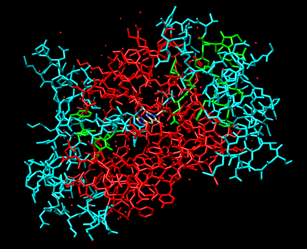
Green fluorescent protein (GFP) is a fluorescent protein discovered in Aequorea Victoria that I’ve found to have many fascinating applications in biological research and clinical scenarios due to its central chromophore within its beta-barrel structure. I conducted a review paper in the ‘exploring proteins’ module focusing on the structure and functions of GFP and emphasised its benefits such as in tumour recognition however, I had a perspective shift upon discovering an article click here that explored previously unknown harmful outcomes of certain GFP strains have on transgenic organisms.

GFP in hematopoietic stem cell tracing

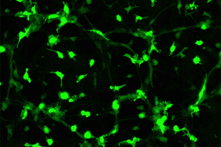
I have a passion for neuroscience and its intricacy including glial helper cells and their functions. Microglia are intriguing as they’re the immune cells of the brain protecting from pathogens and neurodegeneration. Hematopoietic multipotent stem cells produced in the bone marrow are released into the brain and differentiate into microglia cells in response to damage. Through transgenesis, the process by which foreign genetic material is introduced into an organism, GFP has been transduced into hematopoietic stem cells enabling us to track the movement and differentiation patterns as they move to the site of injury.

The use of mutated GFP (enhanced GFPs) combined with an MSCV vector (for stability) in mice chimeras illustrated that upon damage, these stem cells with GFP move past the blood-brain barrier and increase in abundance in the brain. Passage through the blood-brain barrier is rare so this may allow us to create gene therapies against neurological disorders and allow drug delivery similar to that of nanodroplets which I was shown when visiting Southampton General Hospital in which drugs can piggyback into these droplets to the site of injury therefore, something similar could be used using microglia. I think this demonstrates how powerful GFP can be in terms of disease treatments with around 6 million individuals in the US alone being impacted by these types of disorders as shown in this review article the need for this type of research is evident.

The disadvantages of GFP strains

Several new research sources have informed me of the harm GFP expression can cause to transgenic organisms. Several GFP variants show cytotoxic, immunogenetic, and physiological changes to the cardiovascular, urinary tract, and CNS some of which cause death such as in the mice strain alpha-myHC-EGFP which develops dilated cardiomyopathy where the left ventricle of the heart is weakened. The deterioration of GFP as it fluoresces also prevents accurate prolonged tracking. This means the research gathered using GFP in development such as in microglia may not be interpreted correctly. The ASPA Act of 1986 lays out fundamental requirements fulfilled to justify transgenic research projects. GFP is complicated since we still don’t understand which GFP variants cause damage to vertebrates so the rules are less defined.

My views on GFP usage

I think GFP is an incredible protein with the potential to have large impacts on a range of disease treatments and understanding of how the components of the body function and develop however, I also feel the need to emphasise the requirement for research into the different GFP strains and the changes they cause to transgenic animals so that we can properly utilise GFP without the added side effects. GFP in stem cell research has obviously been remarkable allowing us to show the mechanism by which the immune cells develop and create opportunities for treatments that would have a huge impact on a large percentage of the population yet the unsettling amount of unknown knowledge of GFP limits what research I think should be carried out by GFP until further information is obtained.

Will Amniotic fluid stem cells be the future of organoid generation and congenital disease prevention?

Congenital diseases are estimated to be the cause of death for 240,000 newborns within the first 28 days of life and a further 170,000 deaths of children from the ages of 1 month to 5 years old according to statistics generated by the WHO. Identification of these diseases at an early stage Is therefore vital in giving the best chance to apply the most effective treatment whether that be surgery or pharmaceutical therapeutics.

Scientists have had a difficult time isolating tissue-specific stem cells derived from the foetus due to their limited sample obtainability. Collection of foetal tissue has to be done in regulation with ethico-legal restrictions laid out by the Human tissue act of 2004. The extraction of foetal tissue cannot be done past 22 weeks of pregnancy and is usually carried out a post-mortem. The isolation of foetal stem cells has sparked large ethical debates due to the harm caused to the foetus which usually results in the termination of pregnancy. Despite this, these stem cells are crucial in understanding the late-stage development of the foetus and can aid in discovering the severity of cognitive diseases which has a great potential to save many lives.

However the ethical debate may come to an end, a recent study published by ‘Nature Medicine details a groundbreaking revelation into a new method of foetal stem cell isolation through the use of amniotic fluid, the yellow-tinged liquid surrounding a baby during development with the main role of protection. It was found that these foetal stem cells leak into the amniotic fluid during nutrient and urine turnover. This would mean that the direct extraction of stem cells from the foetus will be eluded. The scientists at the Great Ormond Street Hospital and University College London could isolate progenitor cells within the fluid by a technique called fluorescent activated cell sorting or ‘FACS’ along with a range of other techniques to fine-tune the culturing conditions. The cells grew many variations of organoids such as small intestines, lungs, and kidneys in both the progenitor form and epithelia variations. The organoid development will coincide with the development of the organs in the late stage of pregnancy and hence enable us to study the organs in vitro whilst allowing the foetus to continue development. This in-vitro work will enable real-time organoid models representing the current stage of organ development allowing us to recognise and analyse the signs of congenital diseases such as CDH (the condition in which there is a hole formed within the lungs) and hence allow us to come up with personalised pharmaceutical or surgical treatments to counteract the defect by targeting the transcription and genomic expression of the individuals organoid.

This new research has allowed us to understand late-stage development during pregnancy for the first time past the 22-week threshold. The study showed that those who are developing the CDH condition showed a difference in gene and protein expression and hence altered development compared to the healthy organoids. The production of organoids through the foetal stem cells has allowed treatment reflection determining the effectiveness and efficacy of the current treatments available and has allowed the possibility to test new treatments in a less restricted manner.

Currently, the research carried out has led to developments in CDH, TTTS, and MMC treatment however the research showed that within some conditions, improvement has been limited. LUTO stands for ‘lower urinary tract obstruction’. One way this can be treated is by providing a ready supply of amniotic fluid hence removal of the amniotic fluid for generating organoids for further study may be risky hence fewer samples have been obtainable and so the cause of LUTO is still unknown with lack of definite treatments on the horizon due to a variety of isolated cases of the disease.

Overall, I believe this new research has ultimately paved the way to view the late-stage development processes to a further extent than ever before and is an innovative approach that could lead to the almost complete eradication of a whole range of congenital diseases which has the potential to benefit whole spectra of individuals and their families whilst also bypassing one of the most major ethical science debates.

The link to the original nature science paper is here!

3D printed Heart tissue

Just discovered this article in which Stanford university scientists has used a patients own stem cells to develop 3D printed heart tissue. This is a groundbreaking discovery for the patients on the long extensive organ transplant list with the average duration of a waiting time being around 3-5 years this new research can be revolutionary for the treatments and may extend the lifespan of thousands upon thousands of individuals. The method of bio-printing isn’t unique but due to this new method we can now speed up the process by synthesising thousands of cells at a time in so called ‘Organoids’.

Interesting enough, the next step for this research is to develop a way to manufacture multiple Organoids together allowing a more effective way to test new drugs and so this new research has multiple useful applications

check out this video here for more