The University of Southampton

The Stem Cell Saviour

Stem cells have always been an intriguing topic to me, particularly their therapeutic potential. The idea of being able to use one undifferentiated cell to create whole new organs or body parts is fascinating. After our lecture on stem cells and their therapeutic use, I decided to research the different ways that stem cells are currently being used in medicine, and one case in particular stood out.

The Story

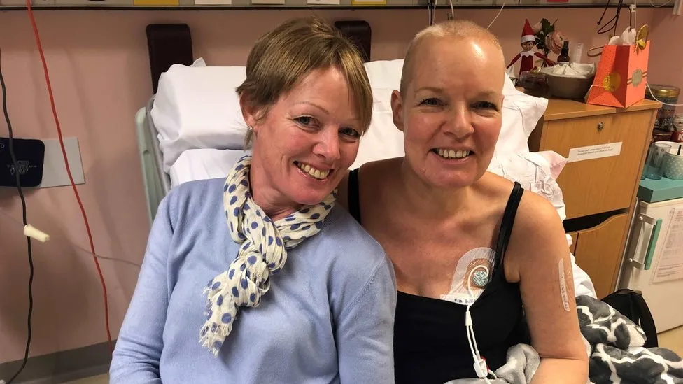
Lesley Calder was diagnosed with acute myeloid leukaemia in 2019. Chemotherapy treatment was unsuccessful for her cancer, and she was left with the only option of a stem cell transplant. Lesley’s three siblings volunteered to be tested, with the slim chance of finding a sibling match (~25%). By some miracle, 2 siblings were full matches and 1 was a half match. Lesley’s sister Annie was chosen to be the donor, and amazingly, Lesley has since made a full recovery.

Using Stem Cells to Treat Cancer

A stem cell is defined as a cell that can self-renew indefinitely and has the capacity to differentiate into many cell types. Their normal function within the body is to replace old, damaged or defective cells to maintain normal tissue function.

Stem cell transplants are used to treat diseases where the bone marrow is damaged or defective, meaning that healthy blood cells can no longer be produced. This is the case in blood cancers (e.g. leukaemia and lymphoma), which primarily affect white blood cells. The loss of blood cells is further exacerbated by intensive cancer treatment (e.g. chemotherapy), which can also damage/destroy healthy cells. The transplantation of stem cells produces new blood cells, and helps to defend against the cancer.

Dr Sonali Smith, M.D. explains the process of using stem cells to treat cancer.

Stem Cell vs. Bone Marrow Transplants

Before this research, I had only heard about bone marrow transplants, and didn’t know stem cell transplants existed, which made me wonder what the difference is. They are essentially the same thing, but differ in the locations where the cells are collected. A stem cell transplant involves collecting stem cells from the bloodstream, which is less invasive than a bone marrow transplant, which involves collecting a person’s bone marrow from within their bone (usually pelvic).

Information from Cancer Research UK says that stem cell transplants are the more common of the two, which I found surprising, considering I hadn’t heard of them. This is because stem cell transplants are: less invasive, easier to perform, have a higher yield of cells and have a quicker blood count recovery.

Dr Scott Bearman, M.D. explains the difference between stem cell and bone marrow transplants.

The Problem & Final Thoughts

Through this research, I have found that over 70% of patients who require a stem cell transplant will not find a compatible donor in their family. Additionally, only 3% of the UK population (and <6% of people in Northern Ireland) are registered to be stem cell donors, making the chances of finding a compatible match even lower. For the majority of patients, like Lesley, a stem cell transplant is their only chance at recovery, and their chances of success are slim.

Lesley’s story prompted her son Max to join the stem cell donor register in hopes of helping others like his mum, and he has already been called upon to donate. By sharing her story, I hope to inspire others to join the register as well, as I will definitely be doing. Anyone aged 17-55 and in good health can sign up here. For more information on stem cell transplants, visit NHS, Cancer Research UK or Leukaemia & Lymphoma Society.

Pre-natal “organoids” – the future of treating congenital disorders?

Congenital disorders (more commonly known as congenital birth defects) contribute to a large portion of paediatric disabilities, and can persist into adult life, if the patient survives that long. The World Health Organisation (WHO) estimates that 240,000 newborns per year die around the world within 28 days of birth due to congenital birth defects. If the affected child survives beyond this stage, a further 170,000 children aged between 1 month and 5 years will die as a result of congenital abnormalities. So, why are these numbers so high? And what can be done to reduce them?

Video from the Centers for Disease Control and Prevention (CDC) explaining congenital birth defects.

As congenital disorders are usually identified after birth, when the abnormality has truly developed and “set in”, treatment aims to improve quality of life rather than cure the patient. Until now!

A “mini kidney” produced from stem cells in the amniotic fluid. Immunofluorescent staining reveals the presence of kidney-specific markers (e.g. GATA3 (distal tubule marker), LTL (proximal tubule marker) and ECAD (apical cilia marker)).

Scientific researchers from University College London (UCL) and Great Ormond Street Hospital (GOSH) have successfully extracted embryo-derived stem cells that are circulating in the amniotic fluid of late-stage pregnancies (i.e. up to 34 weeks), and developed miniature organs (so-called “organoids”) from these cells. Previously, foetal sampling in the UK has only been permitted up to 22 weeks after conception (which is the legal deadline for termination), hindering the ability to study late-stage foetal development. As the stem cells collected by the researchers are sourced from the amniotic fluid, rather than the foetus itself, it allows a ‘bypass’ of the legislation.

The research paper, published this week (March 4th 2024) in Nature Medicine, explains in detail the process of collecting stem cells from the amniotic fluid non-invasively, and culturing epithelial organoids that exhibit features reflecting their tissue of origin (i.e. small intestine, kidney and lung).

Video by Rajamanickam Antonimuthu explaining the process and potential of stem cell-derived ‘organoids’ in prenatal medicine.

This development is extremely exciting for prenatal medicine! The work provides a new opportunity to study late-stage foetal development (something which has not been possible before now), furthering knowledge and understanding. The organoids can also be used to model congenital disorders, extending knowledge on these, particularly in the later stages of development. Researchers now have the potential to develop new methods of diagnosis, prognosis and personalised therapy for congenital disorders, all because of a tiny little organ!

I find this research inspiring and highly interesting, as well as hugely promising for prenatal medicine. Anything that can help reduce the morbidity and mortality of congenital disorders is a huge step in the right direction. The researchers have since expressed their vision to extend the method into the production of organoids from mesenchymal and haematopoietic tissues, allowing the treatment of a wider variety of diseases. I am excited to see how this develops, and, who knows, maybe these tiny organs will be the key to curing congenital birth defects!

Anthrobots – the future of tissue engineering?

I recently came across some research from a team of scientists at Tufts University in Massachusetts, describing their development of ‘anthrobots’. For those like myself, who have never heard of this before, anthrobots are spheres of human tracheal cells that are grown in vitro to form spheroids of a few hundred cells each. The cilia on the outside cells allows the anthrobots to “swim” in patterns, prompting Levin and his team to consider their potential as therapeutic agents.

Anthrobots are spheroids of human tracheal cells (usually a few hundred each) that can be used to deliver therapy.

Levin and his team tested the therapeutic potential of anthrobots to heal a layer of neural tissue that had been damaged by a scratch. They observed that the anthrobots joined together to form a ‘superbot’ – sounds cool, right? What’s even cooler is that after 3 days of incubating the damaged neural tissue with the ‘superbot’, the tissue was completely healed! This surprised the team as this happened without any genetic modification, just the anthrobot’s own functionality. As stated by co-author of the study, Gizem Gumuskaya, it was “not obvious that you’re going to get that kind of response”, prompting the team to think of the wider applications.

Anthrobots come together to form a ‘superbot’, which can then be used therapeutically to repair damaged tissue.