The University of Southampton

Cutting Edge Cuisine : Exploring the Frontier of Lab-Cultivated Meat

Can lab-grown meat be a more ethical food solution?

Recently, a number of my family became meat free. As a lover of chicken nuggets, I was surprised by this, with the scientist in me concerned about the potential drop of protein ingestion. Naturally I was curious about this change, and subsequently gained understanding of how detrimental conventional meat farming is to the environment, the harm to the animals and the cost involved. I recently attended a lecture about tissue engineering and was made aware about how it could be used to grow meat in a laboratory and did more research to determine whether this would be an improved method of meat farming and I believe it is. 

Greenhouse gas emissions from livestock represent 14.5% of emissions

A Global Assessment of Emissions and Mitigation Opportunities, Food And Agriculture Organization of The United Nations (FAO), Rome 2013.

The Science of Tissue Engineering

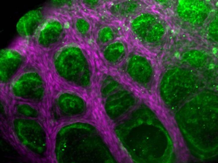
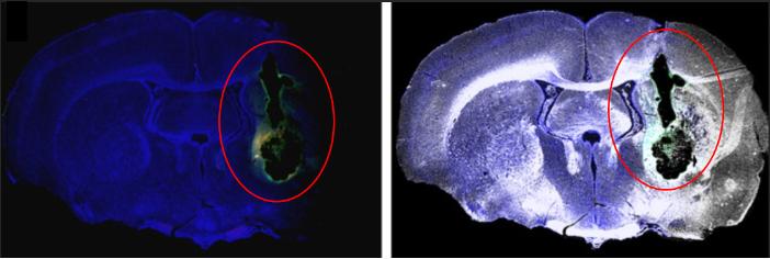
Tissues are made up of cells and an extracellular matrix ; this is anything in a tissue not located inside a cell and is produced by the cells themselves. Tissue engineering involves combining the living component of tissues (cells)  with a scaffolding material for structure and then placing it in a growth media for development. There are a range of scaffolding materials available some are biodegradable and some are not.

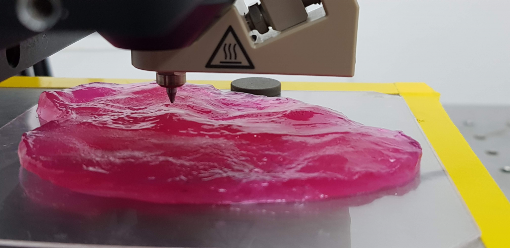
Cooking 101 : How to grow your own meat

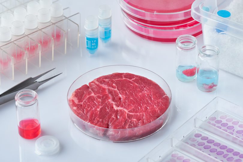
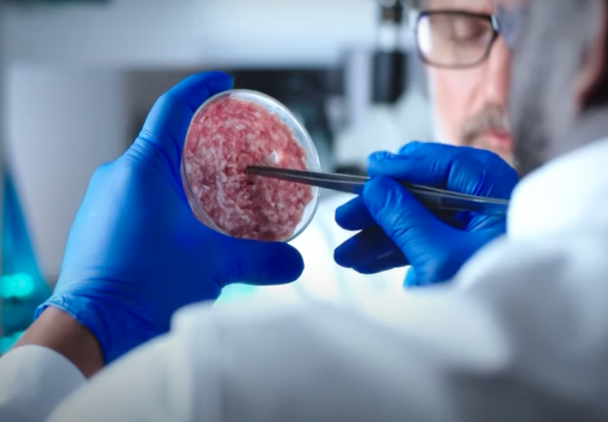
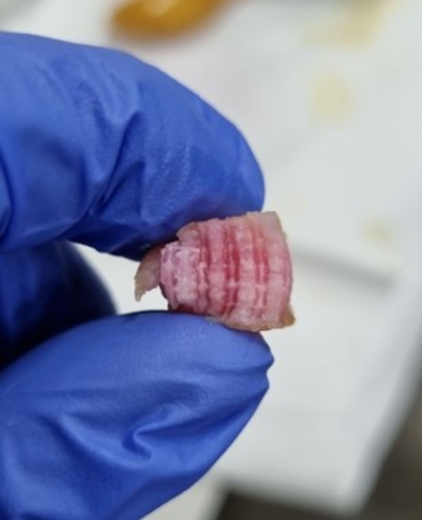
Meat sold in stores is mostly muscle tissue from the animal it’s harvested from with some fat tissue so could be grown in a tissue engineering lab.  

  1. The process begins by extracting cells from an animal, a small sample is taken from an animal under anaesthesia by a trained professional. 
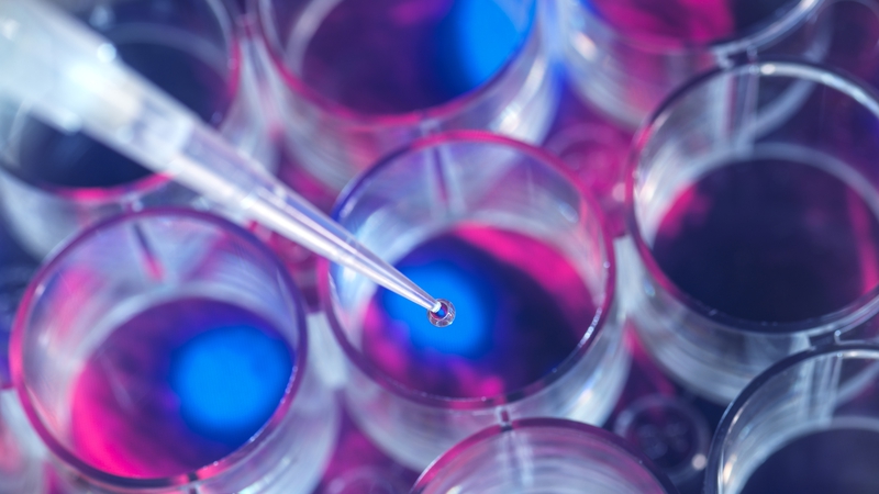
  2. Myosatellite cells (multipoint muscle stem cells) can be extracted and placed in bioreactors with a growth medium that mimics the natural environment the cells would experience as part of an animal (access to oxygen, nutrients and minerals need for differentiation/growth).
  3. Changes in the media composition ,usually cued by addition or activation of the scaffolding material, initiates the differentiation of the muscle stem cells allowing formation of skeletal muscle cells, fat cells and connective tissue.
  4. The differentiated cells are harvested, prepared and packaged before being sent off to be sold in stores.

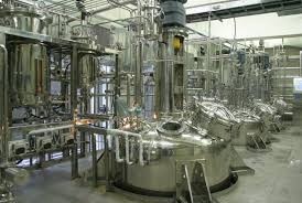
Certain variables like the size of the meat being produced, the growth medium used, the number of stem cells you start with determines the time it takes for this process to occur. Current methods place the timeline between 2-8 weeks which is much faster than rearing an animal from birth for their meat.

Benefits, Limitations and Ethics

Watch this short video to see the benefits of the production process of lab cultivated meat environmentally, to animal welfare and eventually financially.

The main limitations of lab grown meat include:

  • Scaffolding materials are unable to supply oxygen and nutrients to larger tissues like muscle
  • Developing these technologies will be expensive so first generation products might not be affordable to the average household

The main ethical implications are:

  • Animals must still undergo a procedure to extract the cells
  • If the scaffolding material cannot be removed from the end product a hybrid product will be formed as there are no animal based scaffolds currently available.

My Opinion

However, I believe that like me, when people become more aware about the negative effects that large cattle farms have on the environment, how much more cost effective meat production could be as technology advances and ,the biggest reason, that no more animals have to die or be raised solely to be eaten, lab grown meat will rise in popularity and will be properly invested in as an industry.

DIY Artificial Pancreas: a game changer for diabetic care?

During our sensor lectors, the concept of an artificial pancreas was discussed. This stood out to me as my stepmother has diabetes and I had never really considered how having this actually affected her life. It also made me aware that I didn’t even know if she already used an artificial pancreas! Then through researching, I was drawn to the DIY artificial pancreas. I wanted to understand what this was and whether it was the most beneficial option in terms of cost, function and aesthetics.

Individuals with Type 1 diabetes cannot produce insulin to monitor their blood glucose levels. Instead, they inject insulin throughout the day; calculating how much is needed dependent on factors such as what they’ve consumed.

Insulin production for a healthy individual vs an individual with Type 1 diabetes https://www.ndss.com.au/about-diabetes/type-1-diabetes/

What is an artificial pancreas and how does it work?

An artificial pancreas (also known as a closed loop system) can monitor blood glucose levels automatically, calculating and administering the required amount of basal (background) insulin. This is comprised of three components: a continuous glucose monitor (CGM) which uses tiny sensors under the skin that track the blood glucose levels every few minutes, delivering information wirelessly to an insulin infusion pump to administer insulin when the blood glucose levels is not within the target range as well as to a program on a device such as a phone to monitor the readings.

This image demonstrates the components
of the artificial pancreas and how they work.
https://www.bbc.co.uk/news/health-60133358

What is a DIY artificial pancreas and how does it differ?

A DIY artificial pancreas is non-NHS funded but uses similar equipment. It uses a specific app to control your equipment which needs to be compatible with your insulin pump. Guidance is mainly provided through the diabetes community and not the NHS as they have limited knowledge on how these programs work. If you are’ techie’ then you can fine tune this program and ‘train’ the system: making this an advancement on the NHS funded system.

What are the pros and cons?

Now, there are benefits to this: insulin is given automatically when needed which can be ideal especially for young children; allowing a more stress-free experience whilst giving their parents a way to monitor their blood glucose levels throughout the day. It also removes the need to inject which can be beneficial for those who dislike needles. As mentioned, the DIY version can be ‘trained’ to your specific needs so will reduce the energy and brainpower the individual spends on calculating the correct levels. However having said that, because it is technology, this can always malfunction and if given the wrong amount of insulin, it can cause a hypo. The program also requires internet connection where you still need to input your meals as the artificial pancreas can only give basal insulin automatically. In terms of the DIY version, it can be complicated to install and needs updating regularly. Since it needs to be attached to either a belt, a belt loop or a pouch, aesthetically the insulin pump can look bulky under clothing and limit the options of available outfits an individual could wear. This could cause insecurities or just be an added inconvenience.

Video discussing my stepmother’s sensors vs a DIY artificial pancreas

After both points of views it’s clear how life changing a DIY artificial pancreas can be!

Transforming Healthcare: The Application of 3D Printing in Biomedical Engineering

Over the last decade, 3D printing technology has improved drastically, with the main focus from commercial businesses being to reduce their complexity and improve their usability. In contrast, whilst the technology in the medical industry has also improved, complexity has also increased with it. As a Biomedical Electronic Engineer, I am particularly interested in the application of 3D printing within the medical industry.

________________________________________________________________________________________________________________

So, what are the main applications of 3D printing in the medical industry?

Bioprinting

Bioprinting has revolutionised organ transplantation by offering a unique, custom solution. Foreign body rejection can be mitigated using the patient’s own stem cells reducing the need of further surgery. It also eliminates the sometimes agonizing wait time for an organ match – in March 2023, there were around 7000 people waiting for a replacement in the UK alone, so this issue could be avoided entirely. However, the high cost, complexity and ethical implications pose significant challenges, making it inaccessible for some individuals.

An explanation of bioprinting and how it works

Surgery

A 3D printed hip, similar to the one I was shown

3D printing can also be used in surgery to make custom tools for different procedures, and to aid with surgical preparation. One of the most interesting moments for me on this module was visiting the IDS building at the hospital, and learning how scans were used to create a custom hip for a lady requiring a hip replacement. This device helped the surgeons to practice the procedure, whilst also providing the patient with a safe solution. A standard operation would have been difficult and risky due to the level of bone loss; this model helped to replace necrotic (dead) tissue and also bone which had been removed from prior surgeries. After studying modules which covered topics similar to this, it was interesting to see a physical example of a hip replacement, and I found it inspiring that someone could be helped using 3D printing.

Prosthetics

Custom prosthetics can be developed to improve millions of lives – something that motivates me to pursue a career in this field. 3D printers have a massive range of flexibility when it comes to manufacturing, so different designs can be made very easily without the need of additional hardware. I am currently designing a 3D printed prosthetic hand as part of my individual project using my own FDM 3D printer. FDM (Fused Deposition Modelling) is the simplest form of 3D printing; it relies on the layering of molten thermoplastics to produce a 3D object. This project alone has illustrated to me that modern 3D printers have an extremely wide variety of uses at a relatively low cost, with further technological advancements potentially increasing the accessibility of custom prosthetics across the world, helping millions more people in the process.

A timelapse of my own 3D printed hand design using my 3D printer

I personally think that this technology will develop further to the point where any physiological issue can be resolved using something that has been 3D printed – there has been a massive advancement in this field in the last 20 years, so who knows what its capabilities could be in another 20 years!

Overall, 3D printing has provided an efficient solution to complex problems within different industries – especially the medical industry. Different printing methods have contributed to improving countless lives which has inspired many people, including myself, to develop the technology further and hopefully help many more people in the process.

Steady Shoulders: Tailoring Arthroplasty for Epileptic Patients

Epilepsy is a chronic non-communicable disease of the brain characterised by recurrent seizures generally brought on by specific triggers. The most widely accepted description of seizures is tonic-clonic seizures, in which there are brief periods of both tonic (stiffening) and clonic (twitching/jerking) muscle activity but epileptic seizures can manifest in a variety of ways, including absence or loss of consciousness.

My brother is one of the 50 million people worldwide who suffer from this illness and over the previous six years, he has suffered from sporadic, severe tonic-clonic seizures that have irreparably damaged his shoulder, necessitating a shoulder arthroplasty. I didn’t consider the long-term effects of epilepsy as a neurological condition on wider parts of the body until my brother experienced recurring seizures that damaged his shoulder. Upon further research, I have found that shoulder instability is a widely experienced deficit in epileptic patients due to the common risk of dislocation in a tonic-clonic state but can also be caused by the lack of education in seizure management, e.g. restraining someone’s arms during convulsions.

The most common type of instability seen in patients suffering from convulsive seizure conditions is a posterior dislocation alongside skeletal lesions, mainly reverse Hill-Sachs lesions. If the Hill-Sachs lesion covers more than 20% of the humerus head then a surgical procedure is the needed modality of treatment such as a remplissage procedure to fill the indent caused by the lesion or a bone graft to insert additional tissue into the humerus. However if it is not possible to reach clinically defined stability with other procedures or there is simply too much damage to the humerus, scapula and other bone tissues then a full or partial shoulder replacement will be considered. Reverse shoulder replacements are considered when considerable damage has occurred in the rotator cuff so that the replacement will rely on the deltoid to move the arm instead.

Normal shoulder without shoulder replacement
Anatomical shoulder replacement
Reverse shoulder replacement
Explanatory video outlining how a total reverse shoulder replacement is carried out
Explanatory video outlining how a total shoulder replacement is carried out

Some considerations to be addressed when importing an artificial shoulder into an epileptic patient range from seizure control during and post-operation, material compatibility, fracture and wear resistance and potential reoperation risks. In my opinion, the most important qualities needed from the shoulder replacement are high fracture and wear resistance due to the unpredictable nature of seizures making epileptic patients more prone to traumatic injuries. Furthermore, material compatibility is vital in ensuring the arthroplasty fully integrates with the patient and certifies a low risk of shifting during a convulsive seizure.

Material compatibility and fracture and wear-resistance are two factors that seem to work on a pivot as upon further research it seems that materials such as Ti-6Al-4V (titanium aluminium valium) alloys are less wear-resistant but are very effective in osteointegration and hold good osteoconduction properties. Up-and-coming research into joint replacements has already seen impressive benefits in using ceramic and pyrolytic carbons as alternative bearing materials and chemical modifications to polyethelene such as cross-linking, minimizing oxidation, and vitamin E impregnation, have been developed to minimize wear. Improving the bioactivity of orthopedic materials such as mechanically reinforced UHMWPE has been shown as successful and a great candidate to be used for bone replacements (Senra et Al, 2020).

To conclude, shoulder arthroplasty is a safe and beneficial surgical treatment for recurrent shoulder dislocation in seizure-prone epileptic patients (Austin et al, 2023). Upon asking my brother, he has seen a massive improvement in his quality of life thanks to the replacement surgery as he now has a better range of motion, reduced pain and increased stability without having to worry about complications if he dislocates his shoulder during a future seizure. I’d never considered the benefits of replacement body parts, such as shoulders, for patients suffering from neurological conditions but now I can see what is achievable in improving their quality of life when dealing with a debilitating disorder such as epilepsy.

Sources:

https://www.who.int/news-room/fact-sheets/detail/epilepsy

https://www.sciencedirect.com/science/article/pii/S1045452722000803#:~:text=The%20safety%20of%20shoulder%20arthroplasty,cuff%20disruption%2C%20or%20implant%20failure.

https://www.sciencedirect.com/science/article/pii/S1058274602000228?via%3Dihub#bib20

https://my.clevelandclinic.org/health/diseases/24304-hill-sachs-lesion#management-and-treatment

https://pubmed.ncbi.nlm.nih.gov/32890047/#:~:text=The%202%20main%20metal%20alloys,improved%20osseointegration%20and%20osteoconduction%20properties.

Learning Life in Sport with One Less Limb

A backstory…

When I was young, my Granny lost her arm to amputation. Since then, I’ve watched her struggle, relearn, and grasp the basic skills to be able to live an almost normal life again. I’ve always been proud of how she reshaped her life, and since mine revolves so much around sports, I couldn’t imagine coping with a lost limb. This is until I met South African elite para-triathlete, Mhlengi Gwala. He told me about his backstory how he tragically lost his leg and the work he’s done to get to where he is today. I instantly became fascinated with the rehabilitation he endured so I explored deeper into it.

WARNING: Sensitive Content.
A short video on Mhlengi Gwala’s Backstory.

The Road to Recovery

Prediction times for the return to sport after amputation were studied by Matthews et al. I learnt that it’s hard to give an exact period on how long amputation recovery will take, let alone getting back into sports. This is due to factors like what limb was amputated, stump length, the cause, and sport. Think about when you were a baby, you learnt to walk before you learnt to run. The same idea applies to athletes with leg amputations. According to Pam Health, there’re 3 main components to the healing process:

  1. Physical therapy and rehabilitation
Methods of rehabilitation for walking, using bars (left) and a treadmill (right)

During surgery, all unwanted parts of the bone and muscles are removed, and the healthy muscle is reattached to the remaining bone. The amputees need to rebuild strength and flexibility within these muscles and learn to use their prosthetic (if one’s needed) with the help of physiotherapists and rehabilitation teams. 

  1. Managing the risk of complications

Commonly, amputees will run into complications during recovery. These include things like infection, stump pain, phantom pain, etc, which require additional treatment.   

  1. Gaining mobility and independence

Lastly, when the amputee becomes confident enough with the new adjustments, they’re able to live independently. In Mhlengi’s case, he knew that he wanted to get back to racing, and I believe that having something to work towards helped him through recovery and to become a new upcoming para-triathlete.

Is the Price of Sporting Prosthetics Fair?

I mentioned that I was studying biomedical engineering to Mhlengi, who replied “You’re going to be rich, these aren’t cheap”. According to Alan Hutchison, a leg prosthetic can cost up to $60,000. Shocked, I researched why, looking specifically at running prosthetics. A para-athlete with a leg amputation will generally have two different prosthetics because when running or jumping, you’re applying a larger impact force onto your legs. The sporting prosthetic must be able to withstand this, therefore it’s constructed into a curve using carbon fibre and custom-made to fit the stump depending on size, shape, and whether the amputation occurred above or below the knee. Taking these into account, it makes sense why the prosthetic is expensive but it’s still debatable whether this price is too high, especially for someone who’s already faced tragedy. Luckily for Mhlengi, he had many supporters on his side who helped him get to where he is today. 

Summary

I’ve always respected para-athletes for what they do, but I’ve never appreciated the process they endure to be able to do these things. Through this research, I’ve realised that “getting back on your feet” takes a lot of patience, not only through rehab but also in acquiring the prosthetic to begin with, however, it’s amazing what can be achieved when you’re driven by the things you love.

Bioprinting of hair follicles- Is this the solution to hair loss?

Imagine a world where baldness and hair loss is no longer a source of insecurity; a world where those suffering from these can regain their confidence and self-esteem. 

Hair bioprinting isn’t just about aesthetics. It holds promise for treating various hair and scalp-related conditions, such as alopecia, a condition that affects millions worldwide. By harnessing the regenerative potential of cells, this technology may offer hope to those who have been struggling with hair loss for years.

Figure 1: Graphical abstract from Ayaka Nanmo’s study (https://doi.org/10.1016/j.actbio.2022.06.021)

In one specific study, the replication of in-vivo tissue configurations and microenvironments like hair follicle germs has been studied to prepare tissue grafts for hair regenerative medicine. This study suggests an approach for the scalable and automated preparation of highly hair-inductive tissue grafts using a bioprinter.

Figure 2: Graphical description of the process of bioprinting of hair microgels (HMGs) and guide-inserted HMGs (gHMGs).

Hair follicle morphogenesis initiates with the formation of a primordium composed of mesenchymal and epithelial cells, instigating tissue development. Distinguished from other organs, the hair follicle generates the hair follicle germ (HFG) at regular intervals postnatally, perpetually renewing throughout life. In the pursuit of advancing hair regenerative medicine, diverse methodologies have been devised for engineering HFG-like grafts, with one of the most sophisticated methods being the use of bioengineered HFGs. This method entails the compartmentalization of mesenchymal and epithelial cell aggregates to elicit interactions, thereby fostering efficient hair follicle regeneration and establishment of connections with host arrector pili, nerve fibers, and recurring hair cycles upon transplantation. However, this approach may be challenging to scale up to the human setting due to laborious manual preparation steps, especially considering the need for thousands of tissue grafts for a single patient. So, there exists a need for a scalable, highly hair-inductive, and preferably automated approach for HFG preparation.

Exploration into scalable methodologies for preparing mesenchymal and epithelial aggregates has predominantly centered around the concept of cell self-organization. Among these approaches, one notable method entails the seeding of a blend of these cellular components onto a flat substrate. This process gives rise to the formation of spherical aggregates characterized by a mesenchymal core enveloped by an epithelial shell. However, while promising, this technique has encountered a notable challenge: the wide distribution of spheroid sizes. This variability in size could potentially lead to unpredictable outcomes in terms of hair-inducing functionalities. It’s been observed that the size of these spheroids correlates with the number of hairs generated, hinting at the intricacies of the relationship between structure and function in tissue engineering endeavors.

Pairs of collagen droplets housing mouse embryonic mesenchymal and epithelial cells were meticulously positioned adjacent to each other and sequentially underwent gelation. In the subsequent suspension culture, the microgel beads spontaneously contracted, thereby enriching the density of collagen and cells after three days of culture. The contracted microgel beads were termed HMGs or hair microgels. To evaluate their hair-inducing potential, they were transplanted into the dorsal skin of nude mice. gHMGs were fabricated in the same manner, but by placing pairs of collagen droplets on aligned surgical suture guides. The effects of the guides were examined after transplantation into the nude mice.

Fast forward to 2017, where the beauty giant L’OrĂ©al made headlines by partnering with the biotech firm Poietis, renowned for its expertise in crafting 3D models of human tissues, to embark on the groundbreaking endeavor of bioprinting hair follicles. Hair bioprinting is a prime example of interdisciplinary collaboration. It brings together experts from fields like biology, engineering, and medicine, all working together to unlock the potential of this cutting-edge technology.

While the prospect of bioprinting hair follicles holds great promise, further refinement of the process is required and extensive research is needed to ensure its safety and long-term effectiveness. On one hand, the idea of being able to customize hair growth feels like a step towards empowerment, a chance to redefine our appearance according to our own desires. But on the other hand, it raises questions regarding the accessibility and affordability of this technology to a broader demographic that must be addressed, underscoring the importance of making cutting-edge advancements in regenerative medicine inclusive and sustainable. Who will have access to this technology? Will it be reserved for the privileged few, widening the gap between those with the finance and those who don’t? Or will it be made accessible to all, ensuring that advancements in science benefit everyone? It’s not just about innovation; it’s about equity and justice. As we navigate the uncharted territory of bioprinting, we must ensure that it’s not just a luxury for the elite, but a tool for empowerment and inclusivity. Only then can we truly harness its transformative potential for the betterment of all.

Bridge to Recovery: Stem Cell-Studded Scaffolding in Stroke Rehabilitation

Despite 100,000 people experiencing a stroke each year in the UK alone and statistically one in four people over the age of 25 having a stroke in their lifetime, there is currently no clinically effective treatment available to reverse the brain damage caused by strokes.

Last summer my mother had a stroke. Not long ago, this sentence filled me with both sadness and fear of what this actually meant, something not only I was feeling. The road to recovery for stroke patients can be steep and unknown due to the short time treatment can be given within, and the limited options currently available. This led my opinion of stroke research to be pretty negative and views of progress to be at a standstill.

stroke is a life-threatening condition that occurs when the blood supply to parts of the brain is cut off, this lack of blood means no oxygen and nutrients can reach the brain leading to cell death. Strokes are medical emergencies, however, if caught early and treated patients are more likely to make a full recovery with almost no permanent disabilities.

The current treatments available in the UK depend on whether the stroke was ischemic; when blood supply to a region of the brain is blocked, or haemorrhagic; when there is excessive bleeding in parts of the brain. Other factors also include the time since the symptoms began and the presence of any other medical conditions.

Roughly 87% of all strokes are ischemic!

Treatments for an ischemic stroke include both medical procedures and medications. Mainly a medicine called a tissue plasminogen activator is used, which breaks up the blood clots that are restricting flow to the brain.

Whereas, for a haemorrhagic stroke blood pressure medicines are normally prescribed to lower the pressure on blood vessels alongside some form of procedure such as aneurysm clipping, coil embolisation, draining excess fluid and surgery to temporarily remove part of the skull.

Although there are multiple treatments available none are specifically tailored to each patient and can specifically target damaged areas regardless of the time taken to receive care. So, what if there was a way to precisely target damage no matter how long after the initial stroke?

A New Potential Delivery System

In 2018, the University of Minnesota McAlpine research group projects 3D-printed a neuronal spinal cord scaffold.

Recent studies by Baker (2009) and Bible et al. (2009) aimed to develop technology for stem cell delivery to treat brain damage. They successfully attached neural stem cells to scaffold particles, optimised conditions for cell viability, determined the ideal particle size for effective delivery, and used MRI-guided implantation for precise targeting. Observations showed primitive tissue formation within seven days post-implantation, marking significant progress in developing stem cell scaffold matrices for stroke treatment. The study highlighted the potential of stem cell research to enhance transplantation viability using bio-scaffolding.


Researchers implanted neural stem cell-coated polymer particles into rat brains with simulated strokes using MRI-guided needles. Some cells formed a fibrous web within the graft, relying on blood vessel support for long-term survival. Post-mortem MRI images highlighted the grafted area, showing new tissue growth in green fluorescence.

Delving into stroke research has been an eye-opening journey for me, exploring the complex world of neurological damage and potential new treatments. It’s captivating to realise the immense challenges faced by individuals like my mother and other stroke survivors including the impacts on their lives and families, all adding to the urgent need for effective treatments. Investigating the latest developments in stem cell therapy, tissue engineering, and imaging techniques has left me both impressed and inspired for the future of stroke therapeutics.

Harvesting the future : Unveiling the potential of stem cell-cultured meat


Cultured meat tissue created using a 3D printer by Meatech.

Utilising stem cells in 3D printing technology has opened up a massive amount of potential in regenerative medicine and is rapidly improving. 3D printing using stem cells is being used today to revolutionise the food industry by providing an alternative to regular meat!

I believe the industry holds promise and can provide a highly customizable and slaughter free alternative to conventional meats. While also tackling issues like climate change, gas emissions and food waste.

What is 3D-printed meat?

It is a type of cultured or programmable meat replica made from the process of 3D printing using stem cells, attempting to mimic regular farmed meat at a cellular level. Aiming to recreate the taste, smell and texture of conventionally farmed meat.


How is 3D printed-cultured meat made?

While production does require fat and muscle cells from an animal, no slaughtering of livestock is required. The process utilises stem cells that scientists choose depending on the type of meat desired. These cells then undergo a proliferation process, and are bathed in a nutrient dense serum in a climate-controlled bioreactor.

After several weeks the cells will differentiate into fat and muscle cells that form the bio-ink. A robotic arm with a nozzle dispenses the cultured meat filament crafting the meat layer by layer. The arm following instructions using a computer-aided design to form the intended structure.

The product is incubated again allowing the stem cells to further differentiate and mature, the muscle fibres once fully developed will have the right thickness, length and density and after a few more weeks the meat is ready to be cooked and served for consumption!

The video below shows how researchers at Osaka University utilised 3D printing with stem cells isolated from Japanese cattle to make wagyu steak :

FastForward Tech

Why stem cell-cultivated meat?

The food industry is resource intensive and needs large quantities of land and fresh water, With Our world in Data stating that “food production accounts for over a 1/4 of global greenhouses emissions”. By utilising 3D printing and cultured-meat harvests we can address the ethical concerns associated with animal farming, but also decrease agricultural land and water usage, leading to enhanced energy efficiency.

Stem cells are one of the most important cells for any organism due to their ability to turn into other types of cell, and are crucial in advancing the field of synthetic meats.

Professor Hojae Bae and his team at Konkuk university are using immortalised fibroblast cells implanted with two genes to transform them into muscle and fat-laden cells, aided by an optimised 3D-printable hydrogel scaffold to grow cells for printing steaks. The image displays the fat and muscle cells together with microchannels.


The future of 3D-printed meat ?

The cost to produce cultured meat was estimated to be about $700 per kilogram by a lab at Konkuk University, with the average cost of a 3D-food printer being between $1000-$5000 with additional costs for food-grading machines for meat production.

The procedural process can also be difficult, with varying qualities of meat and an entire tissue engineering stage prior to actual printing. The 3D printers specific for production of meat are also in need of further development, to ensure that the food manufacturing process is safe.

With the Singapore Food Agency accepting the world’s first cultivated chicken for sale in Dec 2022, it is only a matter of time before other nations follow. I believe that once these issues are attended to 3D-printed meat will soon play a pivotal role in challenging the environment and ethical issues regarding conventional farmed meat.

Can we build stronger bones?- Exploring the field of 3D printed titanium bone implants

World’s first Titanium chest implant. Credit: HeroX

When we break a bone in our body, we typically require surgery which involves inserting a metal rod into the center of the bone. My younger brother recently broke his leg and had to undergo this exact procedure to allow his leg to heal and support the bone. Seeing what my younger brother had to go through got me interested into the world of medical implants. This led me to titanium bone implants, and how 3D printing is used to make these implants.

The rise of 3D printing has revolutionised the manufacturing of implants, as now it is possible to create personalised implants and prosthetics which help improve the comfort of patients in orthopedic settings. Using 3D printing to make personalised prosthetics can provide flexibility and customisation in orthopedic environments which you don’t get with subtractive manufacturing. I think the use of 3D printing is very beneficial in the medical implant industry as being able to create implants that are personally tailored to individual patients is a development that is crucial to the improvement of implants.

Why titanium?

Titanium is used in implants because it is very biocompatible, which means it is not harmful to any living tissue. Because titanium is very biocompatible, this makes it resistant to corrosion from bodily fluids, which allows it to be more acceptable by the body. Titanium is unique in the sense that it is able to bind with bone and living tissue, which makes it well suited for orthopedic implants. This ability to physically bind with bone allows it to grow into the titanium implant as it heals (a process called osseointegration) gives titanium an edge over other materials.

How does 3D printing make titanium implants?

There are 3 main 3D printing methods which are commonly used to make titanium implants: Direct Energy Deposition, Electron Beam Melting and Selective Laser Melting.

Direct Energy Deposition (DED), is where a high energy source such as a laser is used to melt the titanium powder as it is passed through onto the substrate. The benefit of this method is that it is able to create large parts at a high deposition rate.

Electron Beam Melting involves applying an electron beam to a layer of titanium powder, which is then melted and fused with the previous layer. This method is more suited for smaller, complex parts as it is conducted at a high temperature and in a vacuum which leads to minimal stress on 3D printed parts. Selective Laser Melting, is similar to electron beam melting, but uses a laser to melt and fuse layers of titanium powder.

Video displaying how electron beam melting is applied to make implants. Credit: https://www.youtube.com/watch?v=E7–ZWPVVdQ.

Future outlook of 3D printed bone implants

3D printing has made big strides in the medical implant field as it is at a stage where the potential of this technology can be increased with the improvement in machine capabilities and materials. The emergence of 3D printing can transform the field due the continuous evolution of 3D printed processes which can create innovative implants whilst reducing production costs.

My thoughts

I believe that 3D printed titanium implants can shape the medical implant industry as it is very important to ensure that patients needs are met to ensure a high standard of healthcare and I believe that with the recent advancements that have been made in 3D printing, as well as the benefits that titanium provide for the human body, that we are a step closer into improving medical prosthetics and implants.

Young people, did you hear…?

I think it’s often too easy to forget how many people are affected in one way or another by hearing loss. One of my friends recently got a hearing aid and to see how much her mental health and quality of interactions improved was surprising. I have always lived with hearing that works, and up until fairly recently, I had thought it was only really the elderly and a few younger people who were affected. However learning about hearing loss in this module and from my friend has led me to look further into the use of hearing aids in the younger population.

‘Behind the ear’ hearing aid.

Causes

Looking into acquired hearing loss, I found that most cases were due to either infections or exposure to loud music. Loud music being on this list did not surprise me much as I have had first hand experience of being in places like clubs where I could barely hear the words I was saying let alone anyone else’s. Given how important our hearing is to our everyday lives, I believe that there should be tighter regulations on the volume levels these types of places are allowed to use. A recent study found that more than a billion young adults worldwide are at risk of noise induced hearing loss which is way more than I had ever considered. It seems strange to me that workplaces have noise level regulations, but clubs don’t. How are people meant to preserve their hearing if the legislation is not put in place to protect them? I believe it is very irresponsible for the government to not put a simple limit on these things.

Design

On the topic of hearing assisting devices, it is also important to note that hearing aids have come a long way since they started and are now much more sensitive and discrete. I slightly wonder whether the move towards ‘invisible’ hearing aids is indicating that they are a bad thing that should be hidden away, and I believe these are amazing machines that allow so many people to experience the gift of hearing and should be celebrated instead of being obliged to be hidden away.

Images of five ears with different hearing aids in.
Photos of five different types of hearing aids.

Stigma

I think one of the main problems surrounding young people accessing hearing aids is the stigma attached to them. I know many people think hearing loss and hearing aids are their grandparents’ problems, but this is really harmful to young adults getting the access to devices that would greatly improve their hearing. I feel that this stigma is slowly decreasing but is still a major hurdle for many who would benefit from hearing aids. I think this is in part due to the lack of teaching about them at a young age. I know I didn’t learn anything about hearing aids in the younger population until I came to university and met people my age with them.

Video of a Deaf woman talking about her experiences with having hearing aids.

Given that our hearing is a key sense that doesn’t regenerate, I feel that it is imperative that there is more awareness of the possible causes, and how people can access help. My preconceptions about the people affected by hearing loss have definitely changed, and I am now more aware of the increasing younger population that could be so easily prevented from losing their hearing. This module has led to me making some changes to my life such as wearing earplugs when in noisy environments, and generally being more aware of how loud my environment is.