The University of Southampton

Hip Replacements: A luxury?

As my father prepares for his upcoming hip replacement surgery, I find myself reflecting on his fortunate access to private healthcare. The journey leading up to this decision has been far from straightforward. He has undergone numerous doctor’s appointments, consultations, treatments, and procedures, all aimed at providing relief from his debilitating condition. After exploring all other options, he has ultimately chosen to undergo the hip replacement surgery.

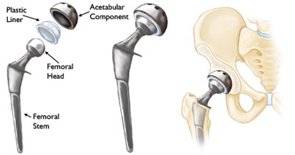
A hip replacement is a surgical procedure where a damaged or diseased hip joint is replaced with an artificial joint made of metal, ceramic, or plastic. By replacing the damaged joint with a prosthetic one, it not only relieves discomfort but also improves overall function, allowing patients to get back to their normal life, something that I am very excited for my dad to get back.

Thankfully, advancements in hip replacement technology have made way for significant improvements in recent years. Enhanced prosthetic materials, including durable ceramics and metals, now closely mimic the natural movement of the hip joint. New surgical techniques and tools such as robotics have all further contributed to these improvements. These innovations have revolutionised patient outcomes, offering swifter rehabilitation and improved quality of life.

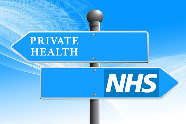
Fortunately, my dad has access to private healthcare through his work and has therefore decided to proceed with the hip replacement privately. Although I am over the moon that my dad can get the care he needs quickly, it has got me thinking about the healthcare accessibility and disparities that exist within our healthcare system.

Not everyone has the luxury of access to private medical insurance. Patients undergoing NHS treatment for their hip replacement may endure lengthy waiting times for surgery. On the NHS website it states that the longest waiting time for a surgery should be around 18 weeks, but this is from the point of referral for the operation, and does not consider the time spent waiting to get the referral, which can be months! This can make the situation worse, diminish quality of life, and impose significant emotional and physical stress on patients and their families.

My dad’s hip replacement surgery is scheduled for next month. Reflecting on healthcare disparities leads me to question whether my dad would have been offered the option for such prompt surgery under different circumstances. I highly doubt it.

I decided to interview my dad to gather insight into his thoughts on his upcoming surgery and to understand the extent to which his hip affects his daily life. Additionally, I spoke with his colleague, Aubery Smith, who underwent a hip replacement surgery through the NHS to gain further perspective (transcript below).

While my father and Aubery Smith both have/ will have their procedures done at the same place, Aubery had to wait “approximately a year” to undergo his hip replacement surgery through the NHS. I cannot imagine enduring the pain he and my father described for such an extended period, and I strongly feel that changes should be made to address this disparity.

Overall, hip replacement surgeries have significantly advanced, offering individuals the opportunity to regain their daily lives pain-free, which is invaluable. However, I believe the healthcare system needs to implement additional measures, such as a reimbursement scheme. This would ensure that individuals in dire need of hip replacement surgery, whose quality of life is diminishing but lack access to private healthcare, can promptly receive the treatment they desperately need. Put yourself in their shoes; wouldn’t you look for timely treatment if faced with a similar situation?

Technology and cochlear implants

In the UK, 12 million people are affected by hearing loss. 80 million are over the age of 60. Over 900,000 people are severely or profoundly deaf, they are unable to hear any speech. During this module, lectures were given on cochlear implants. My interests on these grew so I decided to do some further research. Cochlear implants are used by 12,000 people in the UK.

How do they work?

A cochlear implant is an electronic device that helps with understanding speech and sensing sounds. There are 4 parts: a microphone, a speech processor, a transmitter and receiver-stimulator and an electrode array. The external speech processor captures sound and converts it into digital signals. The digital signals are sent to the internal receiver-stimulator. The signals are converted into electrical energy which is sent to the electrode array that’s inside the cochlea. The electrode array stimulates the auditory nerve by bypassing damaged hair cells. The auditory nerve transmits signals to the brain. These signals are recognised as sound.

Cochlear Implant

Image of CI

Who gets them?

Cochlear implants are given to those that are congenitally deaf, had an infection associated hearing loss such as measles, trauma associated hearing loss like a head injury, age associated hearing loss and people who do not benefit from hearing aids which can be confirmed by specialised hearing tests.

Benefits and challenges

Cochlear implants are given from 6 months old. This is beneficial for children as they are learning to speak and process language. People will be able to hear speech without reading lips including phone calls. They can hear everyday sounds like the noise made when there is a green man at the traffic light. This improves their safety. Cochlear implants help people develop their speech and pronunciation as they can hear their own voice, improving their communication.

There are a few risks, one is meningitis which can occur after implant surgery. This is prevented by vaccinations beforehand. Another, is the results vary depending on the person. Some people’s hearing significant improves while others don’t receive such satisfaction. Cochlear implants are not an instant fix, time is needed for the brain to get used to how they work.

Technology

Cochlear implants are becoming more advanced with technology. Sound processors can be waterproof meaning children and adults can enjoy swimming and even bathe without worrying about any damages. They can be paired with phones through Bluetooth. Sounds can be wirelessly streamed to the processor. An example is the NucleusÂŽ 8 Sound processor which uses improved technology to sense changes in the environment and can adjust listening settings.

NucleusÂŽ 8 Sound processor

Advanced bionics have a cochlear implant called the HiRes Ultra 3D cochlear implant. This device works with technology, making speech clearer and music with better quality sound.

My opinion

Overall, my thoughts on cochlear implants are that they are amazing as they can help deaf people have a better quality of life. I think that they are accessible in the UK due to the NHS and I was surprised to discover that in the US, they can cost between $50,000-100,000. I was appalled by this as many adults and families with children cannot afford such high prices so they won’t get the benefits of cochlear implants. I love that with technology, the external parts of cochlear implants are smaller so people can participate in sports easily. I’ve been to competitions where I saw children with sound processors and I was happy to see them get the same opportunities and experiences as everyone else. I’m excited to see how cochlear implants will further advance.

iPSC’s: The Universal Cure to Human Disease?

Induced Pluripotent Stem Cells (iPSC’s) are a type of stem cell that can be used as a replacement for embryonic stem cells in the research and treatment of human disease. As a Biomedical Science student who has an interest in pharmacology and drug therapies, I believe iPSC’s will lessen our reliance on conventional drugs by targeting diseases at their root cause: cellular dysfunction. The ability to replace damaged cells with new ones provides a novel mechanism for treating diseases, but is it the one size fits all solution to all of humanities ailments?

Introduction

iPSC’s are derived from normal human cells, such as fibroblasts, and are cultured with pluripotency inducing factors such as Oct4/Klf1/Sox2, which are delivered to the cells within a viral vector. This gives the cells the properties of potency and self-renewal that are indicative of pluripotent stem cells. They can then be differentiated into tissues that can be used for drug screening in the lab or inserted into the patient in order to replace missing or damaged cells/tissues. These cells are similar in properties to embryonic stem cells (ESC’s), which are the staple type of stem cell used in regenerative medicine. However due to ethical concerns over the use of ESCs from failed IVF cycles, iPSC’s offer a more ethically sound alternative to be used in regenerative medicine.

What are iPSCs? | I Peace, Inc | Regenerative medicine and drug discovery  through iPSCs

A Simplified diagram of the conversion of somatic cells into iPSC’s

The potential

iPSC’s have the potential to cure a wide range of diseases, from replacing β-cells in the pancreas in Type 1 Diabetes to the replacement of dopaminergic neurones in the brain in Parkinson’s Disease. Diabetes is important to myself as I have many family members who suffer from Type 1 and Type 2 Diabetes, and from interviews with them I found that they struggle with the insulin injection as well as the constant measuring of blood glucose level via a finger prick test. So as a future medical researcher, iPSC’s excite me, as they open an avenue to where diabetes can be cured by replacement of lost β-cells, leading to an eradication of the hampered quality of life people with diabetes must undergo.

Explores the role of stem cells in diabetes treatment.

Explores the role of stem cells in diabetes treatment.

The Issues: Ethical

iPSC’s and their side effects do not line up with Aristotle’s view of ethics as a virtue; as they would not be classified as a high-quality treatment due to their tendency to form tumours, therefore its poor ability to carry out its regenerative function would prove them to not be an effective treatment option. As well as this, in my opinion the use of these cells in a widespread manner may lead to opening pandoras box, where people will look to improve the function of their healthy tissue instead of treating diseased tissue. For example, certain factors may be able to be added to iPSC’s in order to produce muscle tissue with an abnormally high amount of Type IIx muscle fibres, and once transplanted giving them a predisposition to being a successful power athlete such as a sprinter. This in turn will unlock the door to ‘perfect’ humans, a dystopian world with a socioeconomic divide between people who can afford to improve their bodies and those who cannot.

Conclusion

To conclude, iPSC’s are very promising and have a lot of potential for use in regenerative medicine. However, the technology still needs refining and legislations need to be put in place to ensure the technology is not applied outside of disease therapy.

Cautious consideration: The Case for Selective Joint Prosthetic Replacement on the NHS

A patient is admitted with a broken hip from fall and long lie, or even a worn down knee joint, what decides whether they receive the appropriate prosthetic joint replacement surgery? Ultimately, there are professional and ethical obligations to act in the best interests of the patient, but with the NHS suffering from a multitude of economic, political, and social problems that increase the need to be selective in this decision making process, where do surgeons draw the line?

The pillars of consideration to providing this surgery involve: beneficence, assessing whether patient’s lives will get better through surgery; non-maleficence, assessing whether risks and potential complications of surgery are worth it (in both short and long term rehabilitation); autonomy, allowing patients evaluation on the most appropriate treatment including informed consent, ensuring patients are comprehensively aware of all courses of treatment and associated risks and benefits; professionalism, in adhering to the highest standards of practise; justice, providing equitable distribution and access to resources regardless of their demographic.

– find here a youtube clip underlying an orthopaedic surgeons motive for prosthetic joint replacement

Working in trauma and orthopaedics, I often consider whether prosthetic joint replacement is beneficial in the context of neurodegenerative disorders. I have had difficulty in assisting patients that undergo, for instance, hip replacement and day one post-operatively forget they have undergone the surgery. This can involve mobilising prematurely, potentially jeopardizing the integrity of treatment they have received by dislocating or loosening the implant, delaying the healing process, increasing infection or thromboembolic risks, advertently causing more pain and discomfort. This occurs regularly and can be problematic, with excessive treatment and only DOLS (deprivation of liberty safeguards) to prevent postoperative complications. This can traumatise patients who aren’t aware of their circumstances leading to feeling of emotional discomfort, isolation, and lashing out against those trying to help them.

https://www.ncbi.nlm.nih.gov/pmc/articles/PMC9619389/ – find here a comprehensive pubmed publication into the impact of parkinsons disease on the outcomes of total knee replacement

Despite obligations to provide every patient treatment without discrimination, even with ability to use longer term implants like metal on plastic for patients that expect to benefit from the replacement more (those more mobile) compared to shorter term ceramic prosthetics, how far is the surgery beneficial at all considering the cost and compounding factors to those that will not fully understand or appreciate their treatment and could potentially put it in jeopardy?

https://ebjproliancesurgeons.com/blog/understanding-the-different-types-of-hip-implants/ – find here criteria and description for different joint prosthetic media

Could investment for those with neurodegenerative disorder be better spent on research that can prevent higher incidence of falls, reducing the need for surgery at the source? Could it be better invested in mitigating incidence of falls in care by providing more effective infrastructure in place for this? Could investment be better spent elsewhere where the NHS falls behind for those that may truly understand and appreciate their treatment? This decision operates on a fine line and operates on the boundary of what is ethically right.

Ultimately, I have learnt that our obligations to everyone regardless of their demographic or cognitive state are considered equal. Besides, are we not obliged to those suffering from neurodegenerative disorder that were part of the generation that built and paid for the NHS throughout their lives? However, paradoxically, I would consider higher selectivity in providing such surgical treatments as i believe it could be more beneficial elsewhere in patient outcomes and development would lead to better future outcomes for these patients.

https://jamanetwork.com/journals/jamainternalmedicine/fullarticle/414360 – find here a comprehensive study into decision making, procedure, and outcomes of joint surgery in elderly patients with osteoarthritis

Hearing Beyond Silence

This image shows a person with a fitted cochlear implant.

“Blindness separates us from things, but deafness separates us from people.”

Helen keller

Hearing loss currently affects approximately 9 million individuals in England. Projections indicate that by 2035, this number is expected to rise to around 13 million.

A Journey into the World of Cochlear Implants

Attending Nicci Campbell’s workshop on cochlear implants was eye-opening. With a father who battles deafness and relies on hearing aids, I was intrigued to find out more about the candidacy for cochlear implants. Before the workshop, I had a limited understanding of cochlear implants. I mistakenly viewed them as upgraded hearing aids. However, the workshop shattered this misconception. This got me thinking about the gap in public awareness.

The Mechanics of Hearing with a Cochlear Implant

Cochlear implants consist of an external sound processor and an internal implant. The sound processor captures environmental sounds, processes them, and transmits the signal to the implant. Surgically placed under the skin, the implant’s electrodes stimulate the auditory nerve directly, bypassing damaged sensory hair cells in the cochlea. Through auditory training, users learn to interpret the electrical signals.

Are You a Candidate for Cochlear Implants?

Cochlear implants are considered for candidates with severe/profound hearing loss.

  • hearing only sounds that are lounder than 80dB HL at two or move frequencies (500Hz, 1,000Hz, 2,000Hz, 3,000Hz and 4,000Hz) bilaterally
  • AB words test < 50% (for adults)

The day after attending the workshop, my father had a hearing test, eager to delve deeper, I asked him for his audiology reports to review.

This shows my father’s audiograms from 23/02/2024 compared to 20/11/2023 (shown in grey). I compared his audiology report to the degree of hearing loss (displayed on the left). Seeing this made me feel a lot of sympathy for him, despite not qualifying for cochlear implants, his hearing challenges are significant. Specifically:

  • Right Ear: He falls into the severely deaf category for frequencies 3,000Hz and above, and 4,000Hz and above in the profoundly deaf category.
  • Left Ear: Although his overall hearing levels are better, he still experiences severe hearing loss for frequencies 4,000Hz and above.

Clip From a Cochlear Implant User

The workshop featured this video clip of Asgar explaining his experiences. I found that it deeply resonated with me. Asgar’s heartfelt account of his children avoiding conversation with him to evade repeating themselves reminded me of my own childhood -my sister and I would employ the same tactic. I think it’s crucial to recognise that hearing loss not only has a physical impact, but it also effects relationships.

“If I were offered a cochlear implant today, I would prefer not to have one. But that’s not a statement about hearing aids or cochlear implants. It’s about who you are.”

Anonymous

Embracing Silence

Discovering that cochlear implants are not universally embraced initially surprised me. Yet, as I delved deeper, I recognised the profound impact of individual differences. What proves effective for one person may not necessarily be the optimal solution for another.

  • Complex Adjustment Process

Cochlear implants don’t immediately restore hearing, users must learn to interpret the signals. This adjustment takes time and practice.

  • Loss of Residual Hearing

For people with partial hearing loss, cochlear implants can sometimes cause any remaining hearing to be lost.

  • Health Risks

There is a small risk of infection with the surgery.

  • Lifestyle Considerations

Some people may have other strategies for coping, such as sign language and lip reading.

  • Music

While cochlear implants can significantly improve speech, they are not suitable for transmitting the structural features of music.

Pop song:

cochlear implant simulation:

“The cochlear implant is not just a device; it’s a key to a new world of sound and communication”

Dr. Graeme Clark

Awareness

Public awareness of cochlear implants varies widely. Generally, there’s a growing awareness due to increased media coverage and education initiatives. However, misconceptions and stigmas still exist, such as the belief that they “cure” deafness or that they are appropriate for all individuals with hearing loss. Organisations like RNID work on research and public awareness.

Regenerative medicine: The use of bioengineering for neurological recovery

With over 3 billion people in the world living with a neurological condition, the pressure for improvements to be made in this field continues to increase. With my grandfather being one of the many people affected, I am personally intrigued in current and future developments to come. Let’s delve into the interdisciplinary approach of biomedicine and engineering for the recovery of neural diseases.

Neurological disorders

Normal vs Stroke brain scan
Dr Yuranga Weerakkody, & A.prof Frank Gaillard Et Al
Normal vs Alzheimer’s disease brain scan
Australia, Alzheimer’s and Dementia, Health, Diseases and Disorders

The brain and spinal cord are the control centres of the body, sending and receiving sensory and motor information. When this system dysfunctions, it leads to disease. A few examples of neurological disorders are the following:

•Spinal injuries: caused by traumatic damage to the spinal cord, resulting in loss of motor or sensory function, often causing paralysis or impaired mobility.

•Neurodegenerative diseases: include the progressive conditions affecting nerve cells, leading to deterioration of cognitive or motor function, as seen in Alzheimer’s, Parkinson’s and Huntington’s disease.

•A stroke: the sudden interruption of blood supply to the brain, causing rapid loss of brain function, leading to paralysis, speech and vision problems, or death.

The use of biomaterials

The use of human embryonic stem cells for therapeutics to aid brain disorders has come with success but raises ethical questions. It can be argued that the development of biomaterials is a better approach.

Research at Wayne State University in neuronal tissue and cell regeneration to develop the correct biological support network to aid repair.

•For the treatment of spinal injuries, neural grafts and biomaterial scaffolds are employed for functional recovery, physical support and help foster axonal regeneration. Strategies like electrical stimulation and growth factor delivery further aid neuronal regrowth.

•Neurodegenerative diseases prompt the development of 3D brain tissue models, organoids and neural implants to understand disease mechanisms whilst stem cell therapies, including induced pluripotent stem cells (iPSCs), restore neural function.

•Stroke therapies utilise biomaterial scaffolds and injectable hydrogels to promote neuroprotection and enhance functional recovery.

Hydrogels: application in stroke recovery

Hydrogels provide therapeutic benefits through drug delivery, tissue regeneration and wound healing, including direct injection into stroke cavities and forming protective barriers in the brain post-stroke.

MediaNews Group/Boston Herald via Getty Images

Derived from natural or synthetic polymers, they form a 3D structure, absorbing biological fluids within the body. They are flexible and resemble tissues which can be tailored for specific applications. They also play a crucial role in aiding neurological regeneration for stroke patients through multiple mechanisms.

Firstly, they provide a scaffold for cells to adhere to and grow within, facilitating cell migration and tissue regeneration. Secondly, hydrogels can deliver therapeutic agents (growth factors and stem cells), promoting neuronal survival, angiogenesis and modulation of the inflammatory response, essential for recovery. Thirdly, their biocompatibility ensures sustained release of therapeutics without adverse immune reactions. Lastly, their physical properties can mimic the brain’s native tissue environment, fostering appropriate cellular behaviour and functional recovery. In essence, hydrogels offer a versatile platform for neurological regeneration.

Department of Chemical and Biomolecular Engineering, University of California

Things to consider

Concerns linger over longevity, immune rejection, and unforeseen risks of biomedical interventions. Legally, adherence to regulatory frameworks, patent protection and insurance coverage are paramount for ensuring safety and accessibility. Ethically, emphasis lies on informed consent, equitable access, especially in developing nations and addressing social injustices. Who deserves this treatment and why?

My final thoughts

Whilst all sides present a fair argument, I am inclined to take the equitable approach in providing access to these treatments. The choice should be with the patient, neither the law or the socioeconomic circumstances of the individual should intrude on their autonomy or dictate their access to treatment. However in reality this is not always the case.

The long term effects of using biomaterials, particularly hydrogels are a controversial topic. Nevertheless, their use for stroke treatment has shown promising potential, with wider applications to other neurological cases. Caution should be taken with experimental treatments as their long term effects remains to be seen.

Cochlear Implants: Key to an Improved Quality of Life for the Deaf Community?


Hearing – a natural ability we possess and one of the five senses crucial to human experience. Yet, it is often overlooked. Hearing loss is an invisible disability affecting 1-in-6 adults in the UK. This sparked my curiosity about how hearing loss, whether congenital or acquired, impacts quality of life. Especially in terms of its social aspects. This article published in May 2022 states that factors such as “differences in cognitive abilities and age-related changes may exacerbate the problem”.


What Are Cochlear Implants?

A cochlear implant is a small yet complex electronic device designed to provide a sense of sound to individuals who are profoundly deaf or severely hard of hearing. It consists of an external component placed behind the ear and a second component surgically implanted under the skin.

How Do They Work?

As shown by the diagram above, cochlear implants consist of a microphone which captures sound from the surroundings. Detected sounds are sorted and organised by a speech processor. A transmitter and receiver/stimulator then receives signals from the speech processor and transforms them into electrical impulses. These impulses are gathered by an electrode array and transmitted to various sections of the auditory nerve and finally to the brain which recognises the electrical impulses as sound.

This insightful video by Yale Medicine briefly explains how cochlear implants work and that, in effect, they bypass damaged portions of the ear to directly stimulate the cochlear nerve.


Quality of Life

During my research, I found that one aspect to consider within the deaf community is listening effort – referring to the cognitive exertion required to understand and process auditory information. If it is more challenging to hear, it must also be exhausting to communicate. While it may require some acclimating, with the use of cochlear implants, the ability to engage in social settings becomes possible. This can significantly improve quality of life for deaf individuals by reducing the need to be in a constant state of high listening effort.

Individuals with hearing loss often face challenges with employment leading to socioeconomic disparities. Statistics show that the employment rate for those with hearing loss stands at 65%, in contrast to 79% for those without any long-term health issues or disabilities. These figures reflect the situation in the UK – a well-developed country. However, differences in employment rates may be greater in less-developed countries with limited access to healthcare facilities and lack of awareness about hearing loss. A study from Cambridge University found that individuals with cochlear implants reported higher levels of employment and income.

The Downside

Perspectives on deafness vary among individuals. I found this article particularly captivating as it delves into how some may embrace deafness as a cultural identity. The deaf community have developed their own mode of communication over centuries through sign language. To them it is not simply a means of communication but also a part of belonging to a community.

The widespread use of cochlear implants raises concerns within this community about the potential loss of their language and culture. In fact some deaf parents go as far as to request pre-implantation genetic diagnosis (PGD) to ensure their children will be born deaf, and thus take part in their culture and lifestyle.


Conclusion

Cochlear implants overall represent a remarkable advancement in technology for individuals with hearing loss. While they may not be a cure-all, they have the potential to enhance quality of life by restoring access to sound and facilitating better communication. However, it is crucial to recognise that the decision to pursue cochlear implants is deeply personal and not every deaf individual may choose to use one.

This Blog Will Take Your Breath Away

Engineering Bronchial Tissue through the use of Scaffolds to Treat Asthma

Following the lecture on tissue engineering, I felt inspired by the emerging technologies and how these might lead to new therapies. I began to reflect on my own health condition of asthma, and became curious if this relatively-new innovation could be the answer to my future health. Having lived with this condition for almost 21 years, I have always wondered could we, one day, cure asthma entirely?

I began to research the future of tissue engineering in relation to lung conditions, and was particularly interested in Hafeji et al.,’s (2019) paper, Scaffolds for Tissue Engineering of the Bronchi, which has inspired this blog.

So what is Asthma?

Asthma is a common lung condition that causes occasional breathing difficulties. Symptoms include wheezing, a tight-feeling chest, breathlessness and coughing. During an asthma attack, airways become inflamed and the walls of the bronchi constrict, as you can see in the image below. This reduces airflow into the alveoli.

Triggers include infections, allergies, pollution and exercise, but attacks can also occur randomly, which is what I personally tend to find. Asthma affects more than 300 million people world-wide, so finding a cure is essential.

Want to know more about how Asthma works? Check out this video!

So what is Tissue Engineering?

Tissue engineering refers to the assembly of functional constructs that restore, maintain, or improve damaged tissues or whole organs. In this process, cells are selected, cultured and proliferated. For cells to proliferate, a scaffold is required. A large variety of cells are used for regenerative tissue engineering. These include:

  • Allogenic cells = cells harvested from other individuals of the same species, including embryonic and mesenchymal stem cells.
  • Autologous cells = cells harvested from the patient and reintroduced in a secondary site.

Tissue engineering in Asthma

Majority of bronchial tissue engineering studies have involved autologous and allogenic cells, including the use of human bronchial epithelial cells (HBECs) and human bronchial fibroblastic cells (HBFCs). The earliest study of this took place in 1999, where Zhang et al., seeded HBECs into a collagen-scaffold gel after HBFCs were incorporated, resulting in a tissue-engineered bronchial mucosa. This scientific success has lead to the continued study of bronchial disorders such as asthma.

Collagen – the best scaffold?

The role of scaffolds in bronchial tissue engineering is to provide an environment that resembles a native extracellular matrix, promoting proliferation and differentiation. When selecting the material for a bronchial scaffold, biocompatibility and strength must be considered.

The fibre-structure of collagen

Collagen is one of the most abundant structural proteins within tissues, offering high-strength, biocompatibility and even promoting cell proliferation. Even its fibre-structure is ideal -trapping growth factors and allowing HBECs to migrate and attach on the surface, hence allowing visualization under a microscope and study of bronchial disorders. Perfect, right?

Not quite… Collagen as a scaffold has limitations. Often extracted from animal tissues such as pigs or cows, collagen has poor immunogenic properties, posing a risk of carrying diseases.Therefore, collagen must be combined with immunosuppressants which increases cost. This use of animals also creates ethical and religious debate which I worry may limit the demographic that this innovation could target.

The Future

Despite these limitations, the use of bronchial tissue engineering should not be underestimated. I believe tissue-engineered bronchial mucosa acts as an effective three-dimensional model for the further study of this disorder and paves the way for the development of future effective therapeutic interventions, offering hope to millions of people world-wide, including myself.

If, like me, you feel inspired by this, check out Dr Kotton’s current and ongoing research into “rebuilding lungs“. I’m excited to see how this progresses, perhaps making use of tissue-engineered bronchial mucosa as a combined model. The future of asthma treatment looks bright.

Links/Sources:

Breaking Boundaries: Evolution of Skeletal Muscle Tissue Engineering – From Traditional to 3D Printing Innovation

Approximately 45% of the mass of the human adult body is muscle tissue.

Serge Ostrovidov, PhD et al.

Muscles are critical in locomotion, prehension, mastication, ocular movement, and other dynamic activities such as body metabolic regulation. Myopathy, traumatic injury, aggressive malignant tumour extraction, and muscular denervation are the most prevalent clinical indications for therapeutical or cosmetic reconstructive muscle surgery. There are three types of muscle tissue: smooth muscles in the stomach and intestines, cardiac muscle in the heart, and skeletal muscle throughout the body. Skeletal muscle, which we will focus on in this blog, majorly consists of myocytes, the multinucleated contractile muscle fibres.

Methodology of SMTE

While Skeletal muscle tissue engineering (SMTE) has been introduced to the world of biomedical engineering for more than two decades, it remains a significant challenge, with numerous techniques being developed constantly to accomplish this.

The muscle cells are first isolated from the patient or a donor, followed by culturing, where the cells effectively differentiate and develop in the nutrition-rich medium. The muscle tissue is then generated in three methods, which form different specialized muscle cells: myotube formation under controlled topography, cell sheet formation with a hydrogel matrix, and muscle bundle formation. The well-developed and functional tissue is then transplanted back to the patient.

Limitations

This is the traditional way of engineering skeletal muscle tissue, but it still faces some limitations. Though cells could be isolated from the patient, the tissues generated from a donor could trigger immune responses in the recipient’s body, causing rejection and the necessity for immunosuppressive drugs. These medications come with their own risk, like increased susceptibility to infections and organ damage. Moreover, traditional tissue engineering often focuses on preparing damaged muscle tissue rather than promoting regeneration. Thus, the regenerative potential of the SMTE tissue may be insufficient to restore full muscle function, especially in cases of extensive injury or degeneration quantitatively and qualitatively due to ageing. On the other hand, ensuring the long-term functionality of transplanted muscle tissue is also a significant challenge. Without adequate vascularization and support structures, it may encounter degradation or necrosis over time, leading to repeated interventions.

3D Print Muscle Tissue

I came across a piece of news published in 2023 written by Michael M., mentioning the researchers at the Terasaki Institute for Biomedical Innovation have developed a fascinating new technology of 3D printing skeletal muscle tissues with bioink invented by the researchers, which could mimic the natural muscle formation.

Photo credits: Terasaki Institute

The science of muscle 3D printing

The three parts of the bioink are polylactic acid (PLGA) microparticles, myoblast cells, and a hydrogel based on gelatin. The PLGA microparticles sustainably release growth factor-1 (IGF-1), an insulin-like hormone that’s necessary for healthy bone and tissue growth. The production process of muscle tissue is more complex than it looks. Myoblast cells were deemed viable three days after printing and successfully developed into complete synthetic muscle tissue over the next ten days, eventually contracting on their own, just like normal muscle tissues! In order to determine if the cultured tissue was viable in living organisms, the tissue was implanted into mice. The researchers were then able to confirm that the cultured tissue was successfully accepted by the body and combined with the existing muscle tissues.

This video shows 3D-printed myotubes, which eventually form muscle fibres, twitching spontaneously after receiving a dosage of the hormone IGF-1.

Summary

The ability to 3D print muscle tissue represents a significant advance in medicine. Using this technology, muscle mass replacement could be revolutionized by mimicking the natural process of muscle development. Prior to the possibility of treating humans, further clinical studies and testing will be necessary to confirm the safety of this process before the possibility of treating humans. What’s for sure, however, is that 3D printing muscle tissue has a highly promising future in medicine. I, as a biomedical science student, am also looking forward to the further development of skeletal muscle engineering.

References:

  1. Ostrovidov, S., PhD et al. (2014). Skeletal Muscle Tissue Engineering: Methods to Form Skeletal Myotubes and Their Applications. Tissue Engineering Part B Review, 20(5), pp. 403-436. doi: 10.1089/ten.teb.2013.0534

What do you do with good data from bad studies?

Our lecture surrounding ethical dilemmas within the scientific community has really stuck in my brain. Ultimately everyone views situations differently due to individual moral compasses. It also highlighted a key argument: what do you do with good data from bad studies? During the lecture, I concluded that ignoring the data doesn’t undo the crimes committed however it could lead to advances in the medical and scientific field. To see if others shared my views I googled, “unethical studies that generated good data”.

My research revealed it was only in 1945 during the Nuremberg trials that there was an ethical awakening due to prisoner exploitation within concentration camps for medical research. This established the Nuremburg code, which set out to prevent such atrocities from happening again. Unfortunately, studies were conducted before these laws or by people who have no regard for ethical laws now.

Henrietta Lacks

The Henrietta Lacks study is a poignant example of this moral dilemma. Lacks, was in hospital with cervical cancer, unknowingly cancer cell samples were taken and given to researchers. Experiments led to the finding of HeLa cells which became a catalyst for medical advancements in cancer treatments and immunisation.

How do HeLa cells work?

By understanding their function and immortality it is clear they’ve saved countless lives, but the unethical methods by which data was obtained has resulted in controversy.

The lack of consent and financial restitution towards the family outline the ethical complexities of using this data. Surprisingly, Lacks family have started a movement  #HeLa100, promoting the use of HeLa cells, and wish for her legacy to continue. Arguably, there is now familial consent so can the data be classed as ethically sound?

He Jiankui

The He Jiankui gene editing study also interested me, as unlike Henrietta’s story this is more recent, 2018. Jiankui lead unauthorised experimentation on embryos to try and counter HIV, by modifying embryonic genomes to become resistant against HIV – using techniques shown in the diagram. This produced potential benefits within gene editing technology. However rigorous investigations after the research was published led to this statement  â€œHe had defied government bans and conducted the research in pursuit of personal fame and gain” resulting in a sentence of 3 years imprisonment. Fortunately the embryos in question have become three healthy children.

This creates the dilemma – can we use these techniques?

My thoughts

Upon reflection, I think both studies generated data that has led to great scientific discoveries, which fortunately haven’t had negative consequences. It is estimated that HeLa cells have gone on to save over 10 million people worldwide. I think that statement alone argues that we should be using this data even if it was obtained by unethical means. These cells have also contributed towards development of vaccines, preventing the spread of disease and minimizing impact – e.g. the COVID-19 vaccinations.

I also believe that although He Jiankui conducted an unethical study, it’s positive outcome brought about an unbelievable breakthrough in gene editing science. It could create treatments for inherited disorders, that are currently incurable.

Overall, I think the data generated from these studies has led to life changing scientific breakthroughs that if ignored could result in unnecessary deaths and stress on hospitals. But this is just my view, so I would like to put this out there what would you do with good data from bad studies – would you use it?