The University of Southampton

Engineering Replacement Body Parts 2023

UOSM2031

The Alder Hey scandal

I have always been interested in ethical problems; after completing an ethics and philosophy A level I knew I would be interested in the subjects covered in the ethics and law lecture. My mum works for NHS organ and tissue donation, specifically working to improve the way our organ donation system works. This includes the ethical implications that come hand in hand with organ donation. Due to this, I have always been fascinated by ethical issues regarding organ donation.

As soon as I heard about the Alder Hey organ scandal it instilled a great deal of emotion in me, due to the baffling concept of organ removal, retention, and disposal without consent. Especially as a key aspect of the UK’s current organ donation system is based on consent.

What was the Alder Hey scandal?

In 1999 it surfaced that the Alder Hey hospital in Liverpool had been removing various whole organs, hearts, and brains at necropsy from children, without the consent of parents1 . After the inquiry in January 2001, a singular pathologist named Dick van Velzen was charged with committing malpractice.

As outlined in the inquiry the pathologist removed around 850 organs during or after post-mortem and left them in jars, incorrectly processed and uncared for. Many of which were not histologically examined. 

‘systematically stripped of their organs’

Royal Liverpool Children’s Inquiry. Report. London: Stationery Office; 2001. www.rlcinquiry.org.uk/ (accessed 2 February 2001) [Google Scholar]

Reflecting on this information I realised the severity of the Alder Hey scandal. It is difficult to imagine how devastating it would’ve been for parents realising they were denied the opportunity to bury their children whole. For parents processing the unexplainable grief of losing a child, I could only feel pain thinking about how much more difficult the process was made because of the Alder Hey scandal.

What happened as a result of the Alder Hey scandel?

The Alder hey scandal came off the back of the BRI cardiac scandal. Due to the nature and timing of the public release the NHS and government were under a lot of pressure to make a change. The Alder Hey scandal caused a revision of the human tissue act of 19612. The revision claimed to remove any confusion between ‘lack of objection’ and ‘informed consent’ which was where the original confusion lay when collecting organs in the Alder Hey scandal. The department of health and royal college of pathologists should instruct all pathologists that written consent is needed to retain tissue samples and organs. Consent must be gained for each organ retained.

It may never be possible to remedy the pain and suffering of the families at Alder Hey; their legacy, however, must be that activities like those at Alder Hey never occur again.

Bauchner H, Vinci R. What have we learnt from the Alder Hey affair? That monitoring physicians’ performance is necessary to ensure good practice. BMJ. 2001 Feb 10;322(7282):309-10. doi: 10.1136/bmj.322.7282.309. PMID: 11159638; PMCID: PMC1119560.

The revision of this act also brought to light the lack of training for physicians, when talking to and gaining consent from family members. It is not known how many organs and tissue samples collected before the Alder Hey scandal was as a result of proper consent. This showed the need for change. When looking at laws and medical practice and as our technologies and advancements change our laws and practice should change along with them.

The alder hey scandal was specifically eye-opening to me due to my mum’s background in organ donation, along with my idealistic view of our healthcare system. The Alder Hey scandal definitely shook the nation, however I am hopeful that it helped us define consent regarding organ donation. As discussed in the journal, ‘what have we learned from the alder hey affair?’, this part of history will help prevent unethical practice. Teaching us to update medical laws, as we update medical technologies.

References

  1. Royal Liverpool Children’s Inquiry. Report. London: Stationery Office; 2001. www.rlcinquiry.org.uk/ (accessed 2 February 2001) [Google Scholar]
  2. Bauchner H, Vinci R. What have we learnt from the Alder Hey affair? That monitoring physicians’ performance is necessary to ensure good practice. BMJ. 2001 Feb 10;322(7282):309-10. doi: 10.1136/bmj.322.7282.309. PMID: 11159638; PMCID: PMC1119560.

A guide to print your own organs

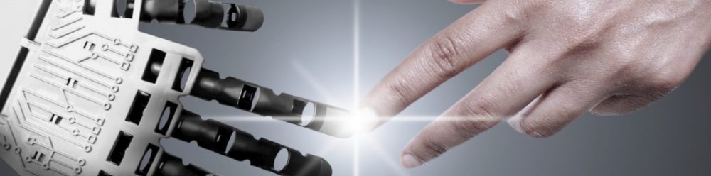
Although the title may seem farfetched, we aren’t as far away from this as it may seem. 3D printing is a technology which is rapidly growing, and is perhaps the answer to many problems within science and medicine.

I became fascinated with the possibilities of tissue engineering after a lecture a few weeks ago, which led me to further research some of the current advances and future possibilities in the field.

It has been 70 years since the first organ transplant, which was a kidney. Since then organ transplant has become a common procedure and has saved many lives. However, there is still some problems associated with it. According to a study by Conor Steward, as of the end of March 2022, there was 4,744 patients on the transplant list in the UK. This long list is costing lives everyday, 3D bioprinting can speed up this process and save lives every day.

So how does this work?

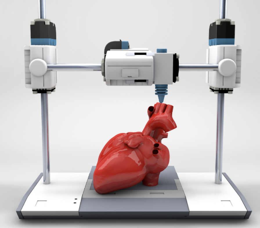
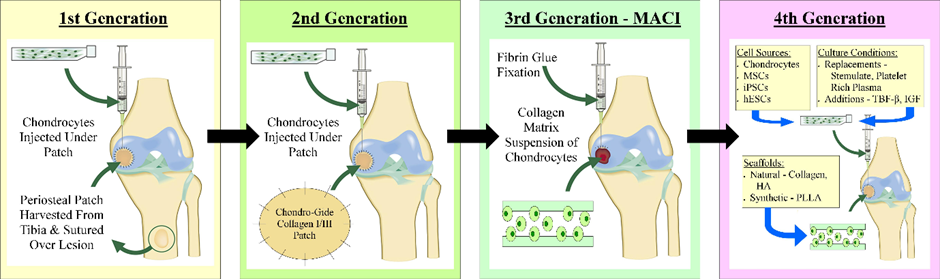
There are three key steps in the process:

  1. Pre-bioprinting – A Digital file is created to input into the printer, telling it what to make. This is often made using MRI and CT scans of the organ you want to print. The cells are then mixed with a bioink, and imaged to ensure they are suitable for the procedure.
  2. Bio-printing – the cell and bioink mixture is placed into the printer, along with the digital file. It is then mixed with a hydrogel as it prints, which is essential in created the structure, as it acts as a scaffold.
  3. Post-printing – to further enhance the structure, the cells are cross linked. This may be via UV light or the application of an ionic solution, depending on the structure.

But where are these cells coming from?

Something that shocked me, whilst learning about tissue engineering, was the range of different sources of cells, and the ethical problems associated with some of them.

The cells may come from a donor, which is known as allogenic cells. There is the possibility here to violate two of the most important laws in biomedical research – confidentiality and consent. In addition to this, one of the problems that strikes me is that it does not reduce the possibility for rejection.

Perhaps one of the most controversial sources is Embryonic stem cells. This involves using embryos to derive stem cells for. However it brings up one of the most pressing questions in biomedical ethics – what is the moral status of an embryo.

However, these ethical issues can be overcome by using stem cells from the patient, that is undergoing the procedure. Cells from your own body are referred to as autogenic cells.

When this comes to mind, you may think of taking stem cells from the patients bone marrow, which is widely used. However I was fascinated to hear how umbilical cord blood can be stored, in the case that a person may need stem cells for any reason in the future. Imagine the possibilities of having your own stem cells, ready for use, in case you ever need them!!

3D bioprinting beyond transplantation

Before researching the use of 3D printing, I thought the only use for these organs was transplantation, however as a scientist, I was fascinated with how they are used for research.

The possibilities of the technology are endless, with studies creating beating hearts, pancreases to cure diabetes, and even a new ear to restore hearing! But in addition to this, we can print tumours or disease models to understand the behaviour of cells!

One study that caught my attention was using 3D printing to create mini tumours, in order to create a more personalised and potent treatment for cancer patients. Scientists at the Seoul National University College of Medicine, formulated a range of bioinks from patients with glioblastomas, which the most common type of brain tumour. A range of chemotherapy and drugs where then tested on these tumours, to allow the scientists to understand how best to treat these tumours. The applications of 3D printing in terms of drug development, is something I am excited to see develop in the future.

Where can it go wrong?

Despite avoiding some the ethical dilemmas associated with xenotransplantation or clinical organ donation, 3D printing brings its own range of moral questions associated with it. In order for the techniques to be readily available, there needs to be tight regulations put in place first.

One problem that strikes me, and many others, is the accessibility of these organs and even personalised medicine. With a technique so new, it will take some time to become widely available, so who gets it first? A problem that has arose a lot within medical ethics, is the use of these products for performance enhancement. This may be particularly prevalent within sports, however it applies to everyone. If you had the money to afford these technologies, you would be able to live a longer life or enhance your quality of life. This sounds great, right? But on the other hand, there is the patient stuck in hospital waiting for an organ transplant, with a life ahead of them with a strict drug regime to avoid the risk of rejection. Is this fair?

In order for the technique to work, particularly to begin with, laws would need to be put in place that the technique should only be used for medical practice, in terms of organ transplant. However personalised treatment plans may be a different issue, as it would be highly beneficial in terms of medical practise, however still separates society.

Personally, talking a utilitarian view point, I believe that the benefits of the techniques are huge, and therefore outweigh any possible socioethical issues that may arise. However as stated above, I don’t think it should be used primarily for performance enhancement, until the technique is widely accessible to everyone, as it creates a larger divide in society than there already is.

Should Euthanasia be legal in the UK?

Trigger Warning – this blog discusses terminal illness and suicide.

If anyone is affected by the topics in this blog, please ring the Samaritans helpline on 116 123.

In the lecture about bioethics and law, one of the areas we focused on was the Nazi’s during WWII and their mass murder or ‘euthanasia’ of people who they deemed ‘unsuitable’.

That made me think about euthanasia and the laws surrounding it. It is legal in Switzerland, and some other European countries and UK citizens often fly there to be euthanised.

My personal opinion is that euthanasia should be legalised in this country with strict legal restrictions and multiple approvals from medical professionals needed.

One of the reasons for my view is because my sister is a nurse who works in ICU in London. She has patients who are on life support machines and have no quality of life yet are kept alive. Some of these were born with such severe conditions, that they have never left the hospital, been able to talk or move, and are also blind and deaf. Is it not crueller to keep them alive, knowing there is no hope for recovery or improvement than to let their parents/medical professionals put them to rest?

In addition, last year my family had to put down our 13-year-old golden retriever. She had cancer and went through surgery and chemotherapy. Unfortunately, the tumour continued to grow, so we stopped treatment and until she became uncomfortable and unhappy. As hard as the decision was to put her down, it was in our home and was the most peaceful and graceful way to die that I could possibly imagine.

Why do we treat our animals with such love and allow them to be euthanised, as the ‘kindest thing’, yet we don’t treat humans with that same compassion? Especially if the human is able to consent?

In 2002, Diane Pretty was diagnosed with motor neurone disease – a chronic and terminal diagnosis. She did not want herself nor her family to suffer through the final stages of the disease. She wanted to end her life peacefully at home. Unfortunately, she was not granted this wish. She went to court to appeal to change the law; however, they did not grant her the right to die. Because of her condition, she had 24-hour care and was unable to commit suicide alone. She wished her husband could assist her with this, however this is illegal in the UK.

Video about the Diane Pretty case
Sharon Johnston and Sue Lawford

How can it be that someone who is able to commit suicide alone is not breaking the law, but someone who needs assistance is unable to? Surely this is discriminatory against those who are disabled.

Last year, Sharon Johnston, who was paralysed but mentally competent, decided that she no longer wanted to live. She therefore chose to be euthanised at Dignitas in Switzerland. Unable to travel alone, a retired NHS worker, Sue Lawford, took her to carry out Sharon’s wishes. Sharon was euthanised but, when Sue got back to the UK she was arrested and questioned on suspicion of attempted murder. She was investigated for six months before being cleared of charges.

It is an incredibly difficult and sensitive topic, and inevitably there will never be a unanimous opinion regarding it. There will undoubtedly be a grey area between assisted suicide, euthanasia, and manslaughter. This potentially puts doctors in a difficult position legally, especially if the patient is unable to consent for themselves. I’m unsure what precise restrictions and requirements should be put in place, but I firmly believe that people who have terminal illnesses or are suffering from incurable conditions should be able to have the choice to end their pain and suffering.  

News article about Sue and Sharon: https://www.bbc.co.uk/news/uk-wales-63599107

My death my decision, a movement that Sue Lawford is a member of: https://www.facebook.com/MyDeathMyDecision/

Your eyes are now obsolete

After doing some research into interfacing electronic signals with the body, I stumbled upon bionic eyes. One device was the ARGUS II, implanted into five patients in England as part of a clinical trial. This article included praise from current ARGUS users Including a man who can now see his grandchildren running around. The articles surrounding the device was overwhelmingly positive until they got more recent then the narrative around ARGUS changed. 

The device is comprised of three modules. An electrode array, video processing unit and a camera. The brain interprets the signals as flashes and the vision created is more akin to an entirely new sense. In this video Ray describes the vision produced as arcs.

The arcs represent the most important parts of the picture. This should be moving objects and edges; the device must accurately pick out the pixels which will convey the most relevant information to the patient. The Video processing unit (VPU) is what does this. It utilizes edge detection algorithms like the matrix mechanics of convolution which I am familiar with from both computer science and quantum mechanics. 

An example of edge detection

The electrical components in the device have several purposes: data and power transfer and biological interfacing. The former is achieved by using magnetic induction. For data transfer the VPU unit encodes signals into radio waves. Which are then converted to tiny alternating currents by a receiver in the implanted region. The data can then be extracted from these currents. Power is transferred by the same physical principle tweaked for this purpose. The final challenge is at the biological interface. The ARGUS II uses micro-electrodes. These define the vision achievable by the device. Advancements in nano-tubes could really improve the quality of the vision created due to physical constraints of wider electrodes.

Despite the quality-of-life improvements seen from most recipients and the prospect of new iterations of devices the device manufacturer Second Sight collapsed and merged with Nano Precision Medical ceasing to develop its ARGUS implant line switching to other projects. The devices had never become profitable. All engineers were laid off and, unlike previously promised, support for the ARGUS models was stopped. Second Sight also failed to inform patients of the collapse. Now users are left to hope their device continues working as normal since replacement parts need to be sourced from the community and many relied upon devices have been rendered not functional by previously routinely fixed hardware issues.

In the EU manufacturers must provide spare parts for a washing machine for 10 years after the appliance has been discontinued. This law was introduced to help limit the environmental impact of E waste and protect consumers. The standard used for washing machines should be the absolute minimum used for implanted medical devices. I believe strongly in the right to repair and choose to repair my own technology, therefore qualified engineers should have access to the parts for device repair long after they have stopped being implanted. 

This is a prime example of our increasing vulnerability in the face of high-tech, smart and connected devices which are proliferating in the healthcare and biomedical sectors.

Elizabeth M Renieris, professor of technology ethics at the University of Notre Dame told the BBC https://www.bbc.co.uk/news/technology-60416058

Many have been left with defunct devices still implanted in their body. I believe the stress and anxiety caused for these people is unforgivable and the law needs to catch up, money and company reputation must stop being placed above patients and transparency.

Commercial Autogenic Cell Therapy Evolution at a Glance.

The lectures I recently received about tissue engineering piqued my interest, specifically with the commercial availability of autogenic cells such as CarticelTM and as I looked further the development of such products was very interesting.

The 4 generations of autogenic chondrocyte implantation (ACI):

First Generation:

A 1st generation ACI procedure is shown in the video below where you can see the harvesting of periosteal tissue from the tibia, suturing of the periosteum into the knee joint, securing with fibrin glue and finally the injection of chondrocytes below this periosteal patch. Genzyme is a company which delivers this service and has reported success rates of 70-90%. Problems with this procedure include overgrowth of the implanted cells which can degrade joint function and cause pain; however this can be easily fixed by the shaving away of excess cartilage. Procedures of this type cost around $40,000, far too expensive for many people, especially in countries without nationalised healthcare such as America where insurance may not cover the procedure.

Second Generation:

Carticel is an example of 2nd generation ACI. A biopsy of cartilage is taken from lesser weight bearing areas so that chondrocyte cells can be isolated and expanded over a period of 4-6 weeks. The expanded cells are reinserted into the damaged joint to form new, healthy cartilage. On their website, Carticel states that their product is intended for the repair of “symptomatic cartilage defects of the femoral condyle caused by acute or repetitive trauma, in patients who have had an inadequate response to a prior arthroscopic or other surgical procedure”. According to the Bioinformant, Carticel autologous chondrocyte implantation costs between $15,000 and $35,000. This cost raises ethical questions because a large subset of people who would benefit from this procedure cannot afford it.

Third Generation:

Spherox is a company which offers 3rd generation ACI with a £10,000 price tag, however the Royal Orthopaedic Hospital (ROH) in Birmingham has provided this procedure and it is now eligible for patients on the NHS according to the ROH website. Spherox works in a different way to Carticel, by taking chondrocytes and producing spheroids of neocartilage composed of expanded autologous chondrocytes and their associated matrix. A sample of healthy tissue is taken from the patient in keyhole surgery and the sample is grown into chondrocyte spheroids. When the spheroids are implanted into the patient’s knee cartilage, they bind to the defective tissue and produce new cartilage tissue. For NHS patients in the UK, Spherox has far fewer ethical concerns regarding cost because the price of the operation is less than the cost caused by such injuries if left untreated to both the NHS and the patient’s quality of life.

The Future:

4th generation ACI therapy has not yet entered mainstream medicine, however various trials are underway. Some research is investigating the role of gene therapy in cartilage repair producing “temporarily and spatially defined delivery of therapeutic molecules to sites of cartilage damage”. According to this paper, the use of elastin as a scaffold is being investigated, as well as the use of a nonviral gene delivery system to allow mesenchymal stem cells to produce osteogenic growth factors.

Empowering Amputees with 3D Printed Prosthetic Limbs: The Future of Accessibility

After attending a lecture by Dr Dickinson and Professor Browne on prosthetic limbs, I found myself intrigued by the manufacturing process behind them. After doing some of my own research, I stumbled upon the recent revolution in 3D printing. In more developed countries such as the United Kingdom and the United States, prosthetics are readily accessible and are usually fitted around 2-6 months after surgery. However, this is far from the case in lower-income countries like Cambodia. It is estimated around 100 million people worldwide require a prosthetic limb; 80 percent of whom do not even have access to this resource. Recent advances in 3D printing technology have made it much easier to construct prosthetic limbs, making it quicker and more affordable as well as being far more tailored to the individual.

A photo showing a donated prosthetic from Guillermo MartĂ­nez to a Kenyan child, synthesised from 3D printing

The benefits of 3D printing

There are several benefits to using 3D printing technology for prosthetic limbs, including:

  1. Customisability: Traditional prosthetics can be difficult and time-consuming to customise, often requiring manual adjustments and moulds. With 3D printing, prosthetic limbs can be designed and printed based on precise measurements of the amputee’s residual limb, resulting in a better fit and greater comfort.
  2. Affordability: Traditional prosthetic limbs can be expensive, with some models costing tens of thousands of dollars. 3D printing technology offers a more affordable alternative, making prosthetics accessible to a wider range of people.
  3. Rapid production: Traditional prosthetics can take several weeks or even months to produce. With 3D printing, prosthetic limbs can be designed and printed in a matter of hours or days, allowing for faster delivery to those in need.
  4. Reduced waste: Traditional prosthetic limbs often require a significant amount of material waste, as moulds must be created and adjusted multiple times. 3D printing eliminates the need for moulds and produces little to no waste, making it a far more environmentally-friendly option.
  5. Innovation: 3D printing technology is constantly evolving, with new materials and designs being developed all the time. This means that prosthetic limbs can continue to improve and become more functional over time, offering even greater benefits to amputees.

Overall, the capability of 3D printing to produce highly tailored prosthetic limbs is one of its main benefits. Contrary to conventional manufacturing methods that call for moulds and human adjustments, 3D printing enables swift and simple production of accurate dimensions and customised designs. As a result, amputees can get prosthetic limbs that match their unique requirements and preferences, enhancing their quality of life entirely.

The drawbacks

While 3D printing technology has many benefits for the production of prosthetic limbs, there are also some potential drawbacks or challenges to consider:

  1. Limited strength and durability: Some 3D printed materials may not be as strong or durable as traditional materials used in prosthetic limbs, which could result in a shorter lifespan or increased risk of breakage.
  2. Quality control: There is currently limited regulation and oversight of 3D printed prosthetic limbs, which could result in lower quality or safety standards compared to traditional prosthetics.
  3. Limited accessibility: While 3D printing technology has the potential to make prosthetic limbs more affordable and accessible, there are still limitations to its widespread availability, particularly in lower-income or developing regions.
  4. Limited design options: While 3D printing technology allows for greater customisability, there may be limitations in terms of available designs or features compared to traditional prosthetic limbs.
  5. Training requirements: As 3D printing technology is still relatively new, there may be a lack of specialised training or expertise in the field of 3D printed prosthetic limbs, which could result in lower quality or inconsistent results.

It is important to consider that many of these challenges are currently being addressed by researchers and developers working in the field of 3D printed prosthetic limbs. As the technology continues to improve, it is likely that many of these issues will be resolved over time.

“The ability to create customised prosthetic limbs through 3D printing technology has revolutionised the field of prosthetics, offering greater accessibility and affordability to those in need.”

Dr. Albert Chi, Medical Director of 3D Printing at Shriners Hospitals for Children

Animal Organ Donors – Is it Worth it?

After attending lectures on stem cells and tissue engineering, I found myself to be very intrigued by chimeras and the diversity capabilities of specific cells. Specifically, the possibility that we could potentially grow human organs in another species, with the intention of organ donation, was something that seemed so unbelievable to me. This provoked me to research further into how close to achieving this we may be, as there is also bound to be ethical issues surrounding this worth discussion. Currently, the NHS weekly statistics reports that 6963 people are currently waiting for organ transplants in the UK. The NHS also reports that 30 in 100 patients experience organ rejection following the implementation of a donated organ. Therefore, I believe it to be an important issue worth researching to find more solutions for.

Pablo Ross (Thursday September 28 2017) working on creating organs from human stem cells that can be grown in pigs and other livestock. Photo Brian Baer

What research is currently being done?

Investigation of this subject lead me to find the research done by Dr. Pablo Ross who has used his extensive veterinary experience, combined with his in-depth knowledge of stem cells to conduct experiments on creating a human-pig chimera. So far, the research and its limited funding, has lead as far as creating chimeras which contain approximately every 1 in 100,000 cells of the pigs being human because pigs and humans are such distant relatives. With this being the extent of current research, it is fair to say we are not yet at a point where human-pig chimeras are able to provide functional help in the medical world. The end goal is that one day we may grow an organ that is fully human or at least made up of enough human cells that it may overcome the prevalent issue of organ rejection.

How ethical is it?

The immediate issue that should be addressed is that testing on animals can not always be seen as entirely ethical as they cannot provide their own informed consent for such procedures and research to take place. It is because of this that humans must make the decision to evaluate if the potential harm to the animals outweighs the benefits of the research or not. The benefits of this line of research, if successful, could potentially lead to a huge reduction in organ rejection, as well as a largely reduced waiting time for all those suffering with organ failure. However, the concept of breeding animals for the purpose of organ donation to humans, which is potentially a very invasive procedure, raises many ethical concerns. My thoughts are that if the research is to be done at all, it may be, in some sense, more fair to use animals who are already being bred with the purpose of being used for their meat as then more of the animal can be used, instead of disregarded.

Simple, yes or no?

So, when I picked this module way back in the summer of 2022, I had no idea that I would become engrossed in a world of engineering. I took biochemistry due to my complete lack of ability in physics, particularly mechanics. However, I soon realised this module ‘Engineering Replacement Body Parts’ dove into far more than the simplistic ignorant view inferred from the course name.

With the main areas of teaching being STEM cells, Prosthesis, Bionics, TISSENG and Ethics and Law I realised I would be receiving an answer to a question I did not know I had.

‘How does the potential of stem cells, engineered tissues and implanted devices in medicine impact the medical field as well as law and ethics in our society?’.

As a Biochemist the area that captured my interest the most was Ethics and Law and the rules and regulations around experimentation. Especially with the current rules changing on gender reassignment surgery for children which has sparked a lot of controversy on whether children can provide consent.

Within my course we have only briefly touched on ethical precautions when conducting experiments, which seems surprising judging how much they govern scientific research. One study that was only touched upon in a Neuroscience seminar I attended was the enforcement of electric shock therapy in the 1960s by Dr. Lauretta Bender. This was a known treatment for psychiatric disorders however she inflicted more than 100 children to shock therapy with the youngest being three years old! Maybe it was because my younger brother had just turned three or the fact that I couldn’t believe that it was not just adults subjected to this treatment. I suddenly thought about a child’s right to autonomy and further what makes someone fit or unfit to give consent. 

Electric shock therapy on a teenage girl in the 1960s by Dr. Lauretta Bender.

It is easy to forget the significance of scientific regulations and ethical boards, as well as how some members of the scientific community only 50 years ago engaged in actions that would now be regarded as atrocities and unbelievable, as shows like Stranger Things and films like Suckerpunch almost trivialise and make medical scandals feel dystopian and alien to us.

The Government currently have legislation on getting informed consent for user research which is shown below directly from the government website:

If this had previously been in place many children would have been saved from the torment and emotional damage they ensued. There are three main views that can be taken when weighing up harm, benefit and autonomy. 

  • Libertarian
  • Paternalistic
  • Utilitarian

All of them would agree that low-risk research where participants are fully informed is an allowable argument. However, both a utilitarian and paternalistic argument would suggest that low risk research where participants do not know they are taking risks is justifiable. This is something I particularly struggled to understand. 

Consent always seemed so black and white, simple yes and no, but when it comes to informed consent, how can a child be fully informed when they aren’t even fully formed?

Biofilms Vs Implants

A lecture by Dr. Alex Dickinson on prosthetic implants highlighted how patient satisfaction rates are a rationale for continued research into implants. I remember learning in Year 2 about how biofilms were one of the major difficulties faced when it came to implants and wanted to look more into what is being done to combat this.  

According to the National Joint Registry in 2013, 34% of knee and hip implants had to be replaced due to infection. Infections of implants result in the patient experiencing similar symptoms to before they had the implant, such as swelling in the area, pain and stiffness.  I can see how frustrating this is for the patients as it must feel like you have gone back to square one. Furthermore, replacing an implant is quite costly and takes up time that could have been used for new patients who require the implant in the first place, not to mention the invasiveness of the treatment.

How does the infection occur ?

In able to understand how treatments are manufactured, I needed to understand how the infection occur in the first place. From my own knowledge, I know that bacteria and other microorganisms enjoy smooth surfaces where they attached on to and  form biofilms. Here they form communities where they ‘live’ which has a constant and stable supply of nutrients to support their survival.

Biofilm Formation

What I did not know is that there are two ways that this infection can occur on the implant. The first being during the surgical procedure – where the bacteria can come from the individuals flora or the operating environment. The second is the microorganisms can be carried by the blood and form a biofilm- this one occurs after the surgery has occurred. In both ways the biofilm maturation occurs over time and it takes some years before it becomes harmful.

By the time the infection is discovered the biofilm has developed so much it becomes difficult for the clinicians to identify what microorganisms that are included in this making it harder to treat them as it is time consuming and requires heavy reach.

What about antibiotics and our immune system ?

Well… multiple studies have shown that biofilms show hight antibiotic resistance due to their formation. When in the biofilm, the bacteria switches off certain genes which could be targeted by these antibiotic rendering them ineffective. Remarkably, one study showed that some bacteria in the biofilm were able express certain phenotypes which would result in the removal of the antibiotics! Due to the lack of blood supply within the implant the immune system is limited.

Current Treatments

So far the most common treatments is DAIR- debridement, antibiotics and implant retention. This procedure involves reopening the implant, washing it out with fluid, removing damage tissue followed by a course of antibiotics , which can vary in duration. The success rates of this treatment vary  due to the heterogeneity of the patients, length of infection and  type of infection. Typically study’s show that DAIR has a higher success rate with acute infection and when the infection has progressed there is an increased chance of the patients reinfection and them requiring are placement  implant. Although there are some success with this treatment , it is still invasive and costly and relies on early detection – which we have seen before is quite difficult.

Future Prospects

I decided to look into research about future therapies or ways to improve the DAIR. I learnt that in order for clinicians to improve the success of DAIR, they are looking into trying to detect the high levels of antibody within the patients so they could be able to intervene at an earlier point. But the invasiveness and time consuming aspects still remain

Antimicrobial peptides have been showing promising results in therapeutics. They already exist in our innate immune system. Their cationic charge allows them kill a range of different microorganisms but not attack the mammalian cells. In addition to this there are other antibiotics such as fluoroquinolones and rifampin penetrate biofilms. This type of treatment ideal but so far researchers have been unable to find a mechanisms for these in vivo.

Copper Vs Bacteria

So if the treatments are lacking, what can be done to prevent this from occurring in the first place? This is where I came across an fascinating paper about looking potential biomaterials which prevent biofilms from forming. The paper looking at what can be done to the biomaterials of the implants to  interfere with the biofilm formation. They conducted a lot of experiments on the metal roughness and tried coating different materials onto the implant. Most notably, the rougher the metal and coating the implants with ions made it harder for the biofilm to form. With this information I began to think that perhaps copper , an ion producing mental could be used to make the implants. Well after reading up on this making copper implants would be impractical but researchers are looking into coating the implants with copper – due to its antimicrobial properties

I was particularly interested in coating the implants with an acylase activity which is turn disrupts quorum -sensing. From studying my course quorum sensing is the way bacterial communicates with each other – so being able to stop that would be detrimental in winning the war against biofilms.

Quorum Sensing

Concluding thoughts

Coming into this, I honestly thought that there was not much hope for resolving biofilm infection of implants. This is mainly because there is not really a reliable , non-invasive treatment for this. However in this case I believe the best way beat biofilm is prevention. The biomaterials look highly promising- although a lot of research has to be done to assure that it doesn’t affect human cells. this is definitely an area that I will keep up to date on as there could be huge developments soon.

Providing better options for prosthetic hand users

A prosthetic hand acts as a substitute limb for those that may be missing one from birth or lost one later in life. There are several types of prosthetic hands, all functioning in their own way and prioritising different aspects for the user. Electrically-powered prosthetic hands offer a range of functions, however are costly. Alternatively, cosmetic prostheses are more affordable but do not provide active function. Because of this, many users ultimately settle with a prosthetic that does not perform to their expectations.

Recently, I watched a documentary that looked into resolving this issue. It showed Masahiro Yoshikawa, a professor from the Faculty of Robotics and Design at the Osaka Institute of Technology, focusing on creating highly functional protheses, while keeping costs low. This is an aspect of prosthetic research that I view as important, as it is essential that prosthetic users are not denied choice because of affordability. Prior to his research, a myoelectric prosthetic was the superior option. This consists of a prosthetic with sensors that detect electrical signals created by muscle movement from the residual limb, triggering a motor. The motor then converts the signal into finger movements, allowing the user to grasp objects. However, this technology is expensive and the prosthetic is heavy, which is not ideal for most individuals.

Offering a low-cost, lightweight, and highly functional prosthetic

Yoshikawa reviewed the manufacturing process for myoelectric prostheses, and figured out that one reason they are so expensive to make is due to the plaster moulds that have to be created prior to making the socket. He concluded that using a 3D printer to create the socket would eliminate the need to produce a mould beforehand, both decreasing manufacturing time and manufacturing cost.

At the core of Yoshikawa’s research is the desire to create prosthetic hands that people want to wear, rather than something people wear because they have no other choice.”

NHK World – Japan: Helping Prosthetic Hand Users Become Choosers

Another improvement Yoshikawa set to develop was the issue of the myoelectric prosthetics malfunctioning after prolonged use. This was because, when users perspirate inside the device, it resulted in a short circuit between electrodes and disrupted the detection of the myoelectric signals. He proposed that, by measuring the height of the muscle as a change in distance, this signal instead could be used to generate motion. More specifically, through using a photoelectric sensor inside the socket, the muscle bulge created by movement of the residual limb will produce a change in distance from the sensor, detected via infrared rays, activating the motor. Consequently, with the sensor not directly resting on the skin and being surrounded by the urethane foam, the issue with perspiration is resolved alongside the benefit of the photoelectric sensor being significantly cheaper than the myoelectric one.

Mechanics of the photoelectric sensor

Since the filming of this documentary, this foundation has been used to create a three-fingered device and a five-fingered device, with realistic options available through the use of a silicone glove.

Different prosthetics created by Yoshikawa

This video shows the three-fingered device in action.

Through researching this topic, it has enabled me to understand the components that need to be considered when developing new prostheses, along with providing equal options for everyone. By developing a lightweight, cost effective, highly functional prosthetic, this has opened up options for individuals who would have previously been limited to a purely cosmetic prosthetic. Hopefully, with the advancement of technology, like what Yoshikawa has demonstrated, a wider range of prosthetic options will be available which users can choose from dependent on their lifestyle.

Check out the link below for the documentary:

https://www3.nhk.or.jp/nhkworld/en/ondemand/video/2015286/