The University of Southampton

The Moral Dilemma of Limb Regeneration: We Have the Technology, But Should We Use It?

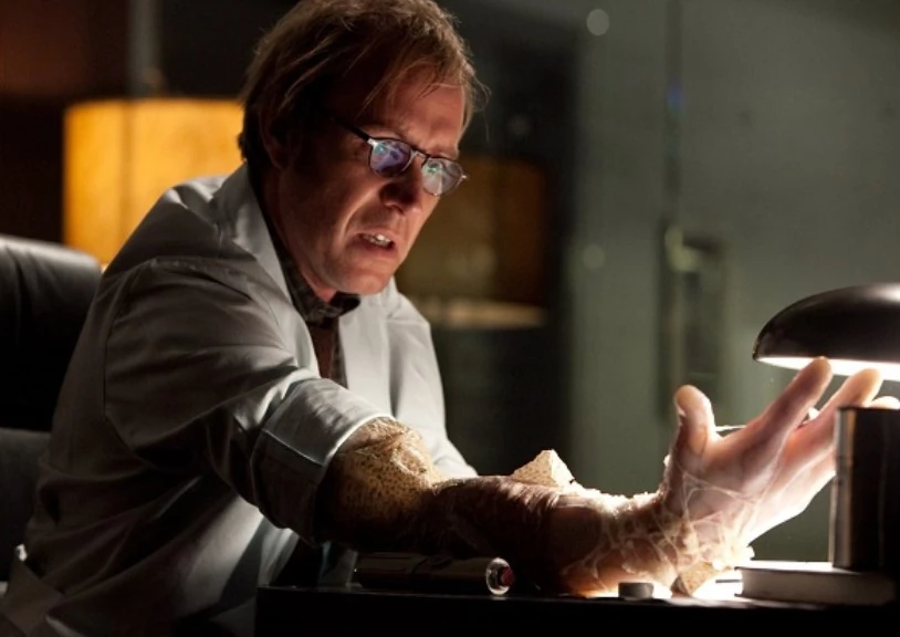
Limb regeneration is a field of research that captured my attention after Dr. Nicholas Evans’ lecture on tissue engineering. The thought of regrowing lost limbs or even organs, was once something only seen in science fiction (like Dr. Conners in The Amazing Spider Man) however thanks to the advancements in tissue engineering and regenerative medicine, it is now becoming a reality. The basic idea of limb regeneration is to stimulate the body’s own regenerative abilities to grow new tissue, bone, muscle, nerves and blood vessels. Many animals are already capable of this such as a salamander.
The process of limb regeneration starts with the formation of a blastema which is a mass of undifferentiated cells capable of enacting growth and regeneration into organs or body parts. The cells have been reprogrammed to become pluripotent. Once the blastema is formed, the cells differentiate into the various types of tissue that make up the limb. They are guided by a complex network of signalling molecules and gene expression patterns. The process is similar to the normal embryonic development of a limb but, it is much faster. While the possibility of regrowing limbs is exciting, it also raises some ethical concerns.

Developing new medical technologies and procedures is expensive and regrowing limbs is no exception! This raises the question of who would have access to this technology? Will it only be made available to the wealthy or to those with good insurance? This could further widen the gap between social classes so is this really necessary?

Another concern is the impact of limb regeneration on the existing prosthetics industry. Prosthetics have come a long way in recent years and many people have benefitted from the advancements in this field. However, if limb regeneration becomes a reality, what happens to the prosthetics industry? Will there still be a need for prosthetics or will they become obsolete? This raises questions about the economic impact of limb regeneration.

Perhaps the most significant ethical concern is the question of whether limb regeneration is even ethical in the first place. Some argue that it is playing God and that scientists and doctors should not be meddling with nature in this way. On the other hand, others argue that it is perfectly ethical as long as it is used for good and not for frivolous reasons like armed forces around the world creating super soldiers.

Despite these ethical concerns, there are many potential benefits to limb regeneration. For example, it could greatly improve the quality of life for amputees, allowing them to regain or gain lost functionality and independence. It could also decrease the use of prosthetics as I said earlier, which can sometimes be uncomfortable and difficult to use. Limb regeneration could also lead to advancements in other fields such as organ regeneration. I believe that the potential benefits exceed the ethical concerns as there are numerous applications of such a process and it would have a significant impact on human health and well-being.

In the end, the decision of whether or not to pursue limb regeneration is a complex one that requires careful consideration both the potential benefits and the ethical concerns. Limb regeneration is an exciting field of research that has the potential to revolutionise medicine and drastically improve the lives of many. With the points I have brought up and with your own opinion, I now ask you, should we use it?

Ethical considerations for 3D bioprinted organs

Following our lecture on tissue engineering, I researched new techniques in tissue engineering and read an article on a woman who had a 3D-printed ear implanted using her cells after being born with a misshaped ear. It’s the first clinical application of 3D bioprinting.

The current goal of researchers is to develop more complex organs like hearts ready for transplant in the next 10 – 20 years. With a shortage of organs to transplant patients and nearly 7000 on a waiting list in the UK alone, the impact 3D bioprinting could have on organ transplantation is immense. It’s crucial progress towards personalized medicine that improves patient outcomes by reducing the risks associated with organ transplants.

As someone interested in working in the pharma industry, I was curious about the issues regarding the ethics of 3D bioprinted organs since all emerging biotechnologies must be ethically assessed before clinical use. Organ transplant currently creates a vast range of ethical dilemmas, so what would happen if we add artificial organs to the mix?

How it works

Although I won’t go into detail on the technique of bioprinting, it’s interesting to learn how 3D printers can print human tissue. Bioprinting relies on bio-ink, comprised of living cells surrounded by hydrogel molecules and acts as a digital file. Once loaded into the cartridge it’s deposited layer by layer of living cells to form 3D human tissue. It’s then crosslinked to become more stable by UV light or ionic solution, allowing it to grow and develop into a functional human organ.

Ethical Issues

The development of 3D bioprinting from laboratory to clinic brings with it established ethical concerns, such as the source of cells. Stems cells are widely used cell source obtained from living or aborted fetuses, raising a bioethical debate to determine their moral status, making their use heavily criticized. Xenogeneic cells are another option as a cell source, although their use has societal and religious concerns, and research has also shown there are psychological issues related to identity for xenotransplant patients. Personally, as a Muslim, I would object to an organ printed using pig cells and would be more inclined to an organ made from my cells. It eliminates the burden of knowing that another person died to save your life, as not all organs can be collected from living donors.

The clinical translation of this technology also brings emerging ethical issues related to personalized medicine to the forefront. Introducing 3D bioprinting into practice requires clinical trials. Generally, trials for 3D bioprinting involve testing patient safety and efficacy of the treatment using randomized placebo-controlled clinical trials e.g. (CAR T cells). Theoretically, it’s possible to test uncertainties related to the risk of harm to a limit, using small transplantable tissue. However, 3D-printed organs made from autologous cells are designed specifically for a patient, making it impossible to extrapolate the results from a randomized clinical trial. Ultimately the patient would have to be their own lab rat.


In addition, due to the invasive nature of the treatment the patient’s right to withdraw as a study participant is challenged. Since the reversibility of organ transplantation is limited, without causing harm to the patient and significantly impacting the chance of alternative treatment. It’s a critical issue for a patient to lose access to better treatment in the future because they participated in an experimental trial. Also, as an experimental therapy, it’s difficult for a healthcare provider to disclose all of the risks associated with treatment. So, even if the patient is conscious and has the capacity they might still not be able to provide informed consent.

Hip replacement – the past, and the future.

Recently I have had the privilege to come to Prof. Douglas Dunlops’ clinic, where I have gained a lot of understanding on orthopaedic surgery. One thing that interested me the most was looking at the development and evolution of hip replacement strategies, and where it can lead us in the future, hence why I have decided to write this blog.

From the beginning

Sir John Charnley was the first to research and develop total hip replacements. He aimed to create a total hip replacement using a synthetic substance between the femur head and the socket, instead of using the natural synovial fluid. After failed attempts with PTFE, Charnley eventually used Ultra-high-molecular-weight polyethylene (UHMWPE) for the first time in 1962. After five years of observing his patients Charnley announced the method as safe, allowing other surgeons to use his patented design and officially making the first functionally total hip replacement. After Charnley’s patent lapsed over 100 kinds of hips were licences, one of them being the EXETER. Its success arose from its tapered stem, allowing it to be easily popped out and replaced, but even-though 92% of them last over 30 years, hip prosthetics still seem to fail.

Less history, more science

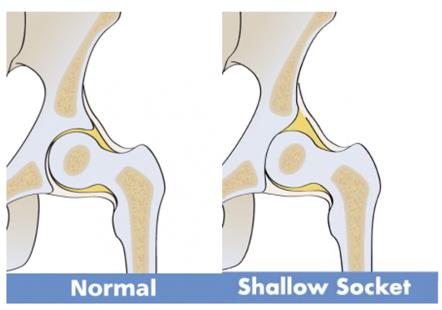
Prof. Douglas Dunlop gave me a lot of insight on all the reasons why hip replacements fail, but one that stood out to be the most and seemed to reoccur was corrosion. The same way the natural femoral head of the hip joint erodes with time, the synthetic joint can cause wear and tear of the cartilage, leading to the formation of a shallow socket and osteolysis. On top of that, physical shearing forces slowly remove the protective film on the metal surface, and any taper interference will corrode the metal further.

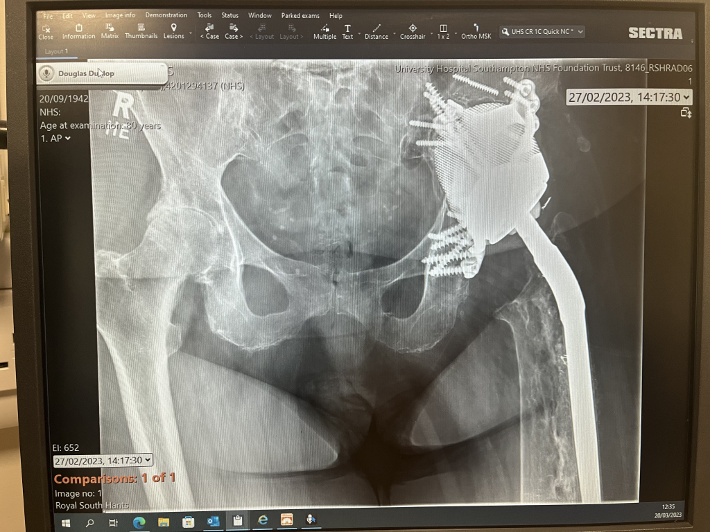
Prof. Dunlop also showed me an x-ray of an elderly patient who had undergone multiple hip replacement surgeries. The shallow socket of the patient caused by hip displasia required for the the prosthetic to be ‘screwed’ into place along with cement loosening. Each time the stem was replaced, the femur had to be reamed, increasing the risk of fracture, prosthetic loosening and infection. This patient had a 3M Capital hip, which prompted the national joint registry due to its poor performance. At the time, the Southampton General hospital was also using a CPT Zimmer brand prosthetic with a fracture rate of 3.4%, giving them a low rating on the registry. In order to overcome this issue, Prof. Dunlop, alongside researchers at Southampton University came up with a solution.

The future of prosthetics

35 years ago, another patient at Southampton General had a similar issue. Her prosthetic migrated and became loose, leaving behind a gap in the bone. In 2003, bone graft from the bone bank was used, but eventually that also failed as screws loosened and the femoral head migrated. It was clear to me that any other attempt at revision would also be unsuccessful, but Prof. Dunlop told me about a new cutting edge technology that has the potential to change prosthesis for ever.

To overcome this issue, 3D custom implants were used, and held in place using stem cells. Not only is the shape of the implant a precise shape for the patient, but the stem cells act as a ‘glue’, allowing for bone formation and encouraging regeneration of the surface layer of damaged cartilage. The removal of old prosthetics may leave behind scar tissue, therefore stem cells may be the only solution to a patient where biology has failed. Upon further reading I have discovered that Dr. Daniel Wiznia of Yale University has developed a similar approach, and deduced that stem cells are a credible strategy and have considerable potential in the future of prosthesis.

Moving away from monoblock stems and exchanging them for a stem with an exchangeable ceramic head seemed to me like a very impactful advancement, but after hearing about the use of 3D printing and stem cells, it has become clear to me that scientists are not done there. It is fascinating to see how the approaches to prosthesis have changed in the last 60 years, and leaves us to think where it can lead us in the future. By writing this blog I aim to show just how fast science is progressing and how successfully scientists are coming up with solutions to clinical problems.

I got the opportunity to listen to Prof. Dunlop talk about his work

The Revolution of 3D Printing in Prosthetic Limbs

Introduction

In recent years, remarkable advancements in the field of prosthetics have been sparked by the development of 3D printing. This technology has revolutionised the way in which we can provide more customisable, affordable, user-friendly prosthetic limbs. One of the biggest advantages of such technology are found in its capability to be accessible to so many where, according to NGO LIMBS, only 5% of almost 40million amputees have access to prosthetic devices(1). This blog aims to expand on the incredible inclusion of 3D printing in the world of prosthetic limbs and how it has the potential to transform lives.

Bone Health Preservation

3D printing allows for custom-fit sockets which have the benefit of using digital scans of patients to develop prosthetics which are more efficient in reducing friction and evenly distributing weight across the limb. This means that bone is less likely to be lost as a result of the prosthetic, which can sometimes occur when the prosthetic socket is not an accurate enough fit. Another benefit of this technology, is its capacity for easy adjustments and remodelling to ensure a consistently well-fitted prosthetic, decreasing the chance of any complications leading to disuse and resultant bone health consequences. Additionally, the 3D printing of prosthetics often uses thermoplastics which are much lighter weight and therefore place much less pressure on the bones. Resultantly, bone density is more likely to be preserved and fractures are more likely to be avoided, aiding the maintenance of bone strength.

Disadvantages and Solutions

Although there are benefits to the use of thermoplastics in prosthetics, they are a less strong or durable option when compared with other materials used in prosthetics, such as carbon fibre. This means that it generally has a shorter lifespan and is more likely to need replacing and therefore may be significantly less suitable for use in lower-limb prosthetics. This may particularly be the case for patients who have more active, high-impact lifestyles, or those with higher body weights.

Therefore, a more suitable use for 3D printing technology in prosthetics, as proven thus far, may be in upper-body limbs and in children. This combats complications of exerting too much pressure on the lighter-weight thermoplastic, and enhances the technology’s potential for durability. This may be a particularly beneficial advance in technology for children and growing adults. This is due to, as before mentioned, the precise adjustability of 3D printing. Additionally, it is a far cheaper option for prosthetics as traditional prosthetics can cost thousands to replace as children are out-growing them. The organisation, e-NABLE began a project whereby many constructed and donated 3D printed prosthetic hands to children for free to aid research in the area.

Summary

The amazing affordability and accessibility provides an abundance of potential for 3D printing in the field of prosthetics. Although there may currently be limitations as to what field of prosthetics this technology might be best suited to, there is hope that technological advances in thermoplastics or finding more suitable materials may lead 3D printing to be the future of prosthetics.

(1) – https://www.sculpteo.com/en/3d-learning-hub/applications-of-3d-printing/3d-printed-prosthetics/

How 3D Bioprinting will revolutionise Therapeutics

A lecture on tissue engineering introduced me to the concept of Bioprinting. Although I am fully aware of the idea of 3D printing. I wanted to look more into how biological materials are incorporated into 3D printing to create tools that can be utilised within our bodies. Bioprinting is ‘the use of 3D technology with materials that incorporate living cells’. They are most commonly known for organ growth to battle the organ transplant crisis. But did you know that they are also being used to improve therapeutics, specifically drug delivery? I was particularly drawn to this as it shows potential in making medicine more personalised – a topic that I have always had an interest in.

3D Bioprinting

Bioprinting and its biocompatibility

There are different types of 3D Bioprinting techniques such as stereolithography and fused deposition modelling. However, bioprinting with bioinks is able to produce structures that obtain many properties that can be used to  develop structures that can be efficient  for drug delivery.

Bioinks are hydrogels that can be made up from substances such as gelatin, alginate and hyaluronic acid – materials that are highly compatible within the body. The composition of the hydrogels can vary and as a result they can be modified so  the cross linkage  of the polymers can be made reversible or permanent depending on how the materials are treated physically or chemically. These are referred to as methacrylate hydrogels and the cross linkage can be used as a tool to be used to control the release of the drugs within the body. The bioinks can also incorporate synthetic polymers which can be used to further modify their properties such a degradation, increasing the biocompatibility.

Potential therapeutic uses

  • Drugs

The main issue in drug therapeutics is the distribution of the drug itself within the body. In addition to this due to individual differences, a set dosage doesn’t work for everyone and a patient may not necessarily take their medication correctly. (Martinez et al., 2017) was able to print drug-loaded hydrogel with water which was able to release the drug at rates that were dependent on the water content. A delivery system, that would combat the improper use of medication, release the drug at the correct site of absorption, and could be further modified to be dose specific for each patient.  

With further reading, I discovered that tuberculosis required two drugs for it to be treated, rifampicin and isoniazid. Multiple studies have shown that these two drugs can negatively impact the effectiveness of the other and they are also required to be absorbed in different areas within the body for effective treatment. (Genina et al., 2017) designed an oral dual-compartmental dosage (dcDU) unit via 3D printing and hot melt extrusion

Design of the Dual – compartmental dosage unit

The dcDU  was tested in vitro and in vivo using rats, where drug release was analysed and it was noted that sealing and the compartments improved the drug release of each drug.  This study particularly opened my eyes to the amazing things that could be achieved with bioprinting, it was here when I begun to realise how much of a big impact on therapeutics that bioprinting would be. An infection that could require a 2-week course of 2 different antibiotics would be reduced to one oral dosage.

  • Hormones

Like any other scientist, I began to work what else could we dispense and came across (Tappa et al., 2017). Intrauterine devices are the most common form of long term birth control. Although they are very efficient they are not necessarily personalised to the individual. Polycaprolactone pellets (PCL) were first coated with hormones and infused with silicone oil. Here is prime example of how synthetic polymers are used as the PCL allows for degradation and has a low melting temperature that will not interfere with the high degradation temperatures of the hormone. They then used this to print different 3D hormone-releasing IUDs which suggested promising uses in gynaecology applications.

Six different biodegradable hormone releasing IUDs that were bioprinted – different shapes could be printed that could be more suited to the patients dimensions making the implant more comfortable
  • Growth factors

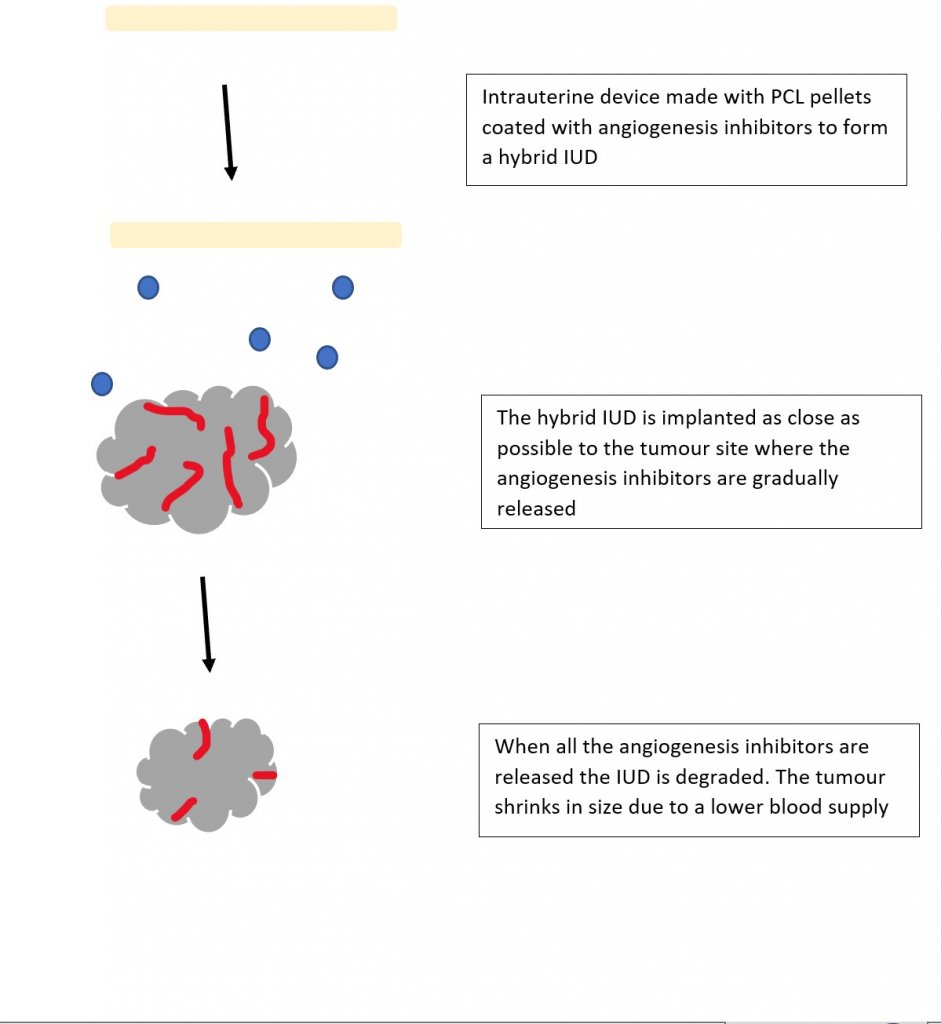
I am very familiar with the growth factor VEGF and its role in bone vasculature, so I really enjoyed (Poldervaart et al., 2014) paper on designing gelatin microparticles which had a controlled release of VEGF. Migration assays showed that the VEGF release from these microparticles lasted up to 3 weeks and was able to induce migration on endothelial progenitor cells. I began to wonder if 3D printing could be used for the potential treatment of bone cancer. What if we combined the biodegradable IUD concept but instead of hormones it contained angiogenesis inhibitors?

Organoids and chip in organs

To test my IUD hybrid idea, I could also utilise bioprinting to create organoids which are tiny 3D tissue culture that allows us to test the effects of infectious diseases and drug therapies. Organoids are rarely studied alone and are often in association with chip-in-organs, which replicate the organ environment so the results are more accurate to the behaviours that would be seen within the body.

Emphasising the importance of chip-in-organs

Future Prospects

A lot of this research is very new and perhaps the two most obvious next steps for Bioprinting are refining the bioprinting mechanism itself and clinical trials, which some predict could occur within the decade. Most importantly, the ethical side of this needs to be discussed. Currently the FDA does not have an official definition for bioprinting, making it harder to set laws and guidelines for it. There is also a bit of worry about potential ‘black market’ uses of these resources – where do we draw the line? I could imagine that people could use this for recreational drugs such as nicotine for example or body builders could use it to increase their testosterone levels

However, the bioprinting world is such a new concept in the world of science with very promising results that could completely change the way we use therapeutics entirely and to be honest I am very excited to witness this.

From stealing organs to growing organs from pigs: the lengths humans will go to survive.

As a registered organ donor before the opt-out system, I could not fathom why people would not want to donate their organs. It baffled me, and yet in 2019, I was in a debating competition where I had to oppose the idea of organ donation as an opt-out system – my point was simple. Surely the argument that this is a violation of a donor’s autonomy and implies given consent, would always triumph over the utilitarian view, that it should be the greatest good for the greatest number, right?

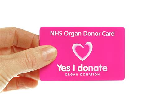
The original donor card required for the opt-in system.

In 2020, the UK introduced a new law, the ‘opt-out’ system. Many believed this diminished a donor’s autonomy, yet, by definition, there is still a choice in opting out. The new law made sense to me, when around 6,945 people are currently awaiting a transplant in England, I found it hard to understand the counter-argument.

Dr. Jon Dawson covered the ideas of organ donation and autonomy throughout his lectures. I saw a wide range of views in one group of students of a similar age and with the same privilege of higher education. Dr. Dawson put forward the Alder Hey case, where about 850 organs were being harvested after death without any form of consent from either the patients or guardians.

A video briefly describing the Alder Hey Scandal.

There was a large consensus that this was not okay from the class, the lack of adequate consent when removing organs and tissue from patients was barbaric, nonetheless arguments can be made that people uneducated in the opt-out system are therefore giving ill-formed consent.

This case made me think of the book Never Let Me Go by Kazuo Ishiguro, where clones are created for the purpose of organ donation, and once they have donated around three of their organs, their short lives are over. Although this dystopian novel seems far stretched, the premise behind it still stands. Especially as since 2015, advancements in tissue engineering has shown animals as a viable surrogate for growing organs.

In 2019, Hiromitsu Nakauchi had the first approved experiments to allow a human-animal hybrid to grow fully. This sounds like some werewolf science-fiction, Morbius esc (awful movie); however, this could be the key to the current organ shortage. In an ideal world, we could grow the organ required at the drop of a hat- but here is a scientific solution where we could grow organs within animals and harvest them without invasive surgery on humans, or an ethical debate of autonomy.

Now, PETA and animal rebellion may be opposed to this idea, but I think animals will forever hold a place in scientific research, so could this be a legal viable solution? What do you think?

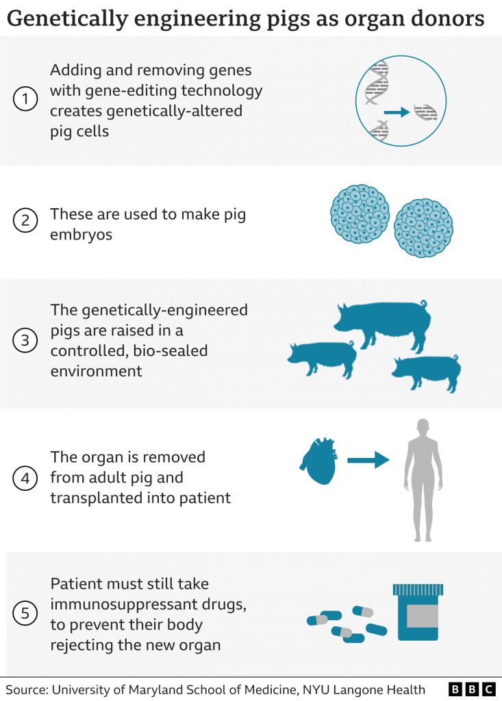
Well, in January 2022, the first pig to human transplant was done; a genetically engineered pig’s heart was harvested and placed into a patient. Although this required a lot of medication and extra resources, the heart did work prolonging the patients life for two months. This was a major breakthrough- can we now combat our organ shortage through animals?

Whether genetically modified pigs or human-animal hybrids hold the future in organ donation, ethics must be considered- we can not return to being so desperate as to take organs without consent. The podcast below is an informal discussion on organ donations with the opinions of two biochemists discussing frankly the possible future of organs, ethics and consent for further insight into this medical and ethical minefield.

https://open.spotify.com/episode/4inRLDaXEcnROyTexaC3WQ?si=7988ff562cb04df4
STEM Sundays a podcast by Lara Etheridge and Yasmin Yardley discussing organ donations, ethics and movies.

Apocalyptic Prosthetics: How realistic are prosthetics in tv shows?

Recently I have been watching the tv show Alice in Borderland, which is set in an abandoned, seemingly post-apocalyptic Tokyo, where players have to complete dangerous to survive. One character, Akane Heiya, has a transtibial amputation due to an injury from a game and uses a running blade. I decided to look into how realistic her prosthetic is.

Obtaining the prosthesis

Since I was watching the show around the time I attended the lectures on prosthetics, it made me wonder where Akane was able to get a prosthesis, since I learnt from the lectures that the sockets are usually made custom for the shape of the residual limb. From further research, I also found that the residual limb often changes shape in the healing process and loses volume, and liners are worn between the limb and the socket to protect the skin which are changed daily (1). Akane wears bandages between her skin and the prosthetic, which could be changed regularly, but I don’t think it would have been feasible for her to find a fitting socket.

Akane uses bandages to protect her skin.

I did not think it would be realistic for her to find a running blade in an abandoned city. From looking into the availability of running blades I came across the Blade Library. This is a facility near the Toyosu Running Stadium in Tokyo which allows amputees to try running blades (2). There are 24 blades available to rent, as well as fitting workshops and and monthly runs to learn how to use blades (3). In the context of the show, it may have been possible for Akane to find a fitting blade here but would probably require prior knowledge of its location. However, I do think that the concept of a blade library is a really good resource to make running more accessible to amputees.

More about the Blade Library in Tokyo.

Using the prosthesis

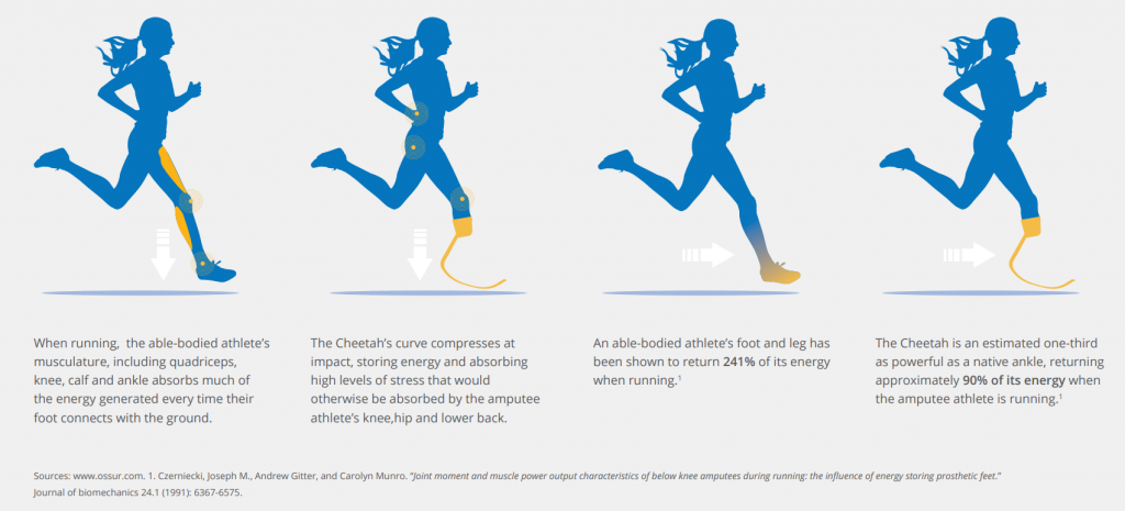
The prosthetic that Akane uses is a Flex-Foot Cheetah design (4). It is a carbon fibre running blade that is attached to the socket with a pylon connector (5). The curved blade stores the potential energy from the user stepping down into the blade and uses this energy to propel them forward (5). Akane uses a C-shaped blade, which is better for long-distance running than the J-shaped sprinting blades (6).

How a running blade works in comparison to an able-bodied athlete.

Although running blades are lightweight and durable, they are not made to be used as an everyday leg as Akane does. Running applies pressures to the residual limb in different areas than walking does, so wearing the wrong prosthetic for either activity can cause discomfort (7). They are usually slightly longer than the standard prosthetic to account for impact when running, and require slightly different alignment, so the knee has to remain bent whilst walking or standing (7).

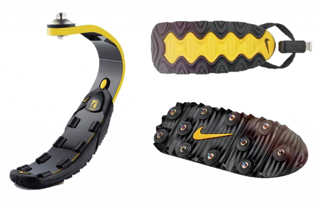
Akane travel across different terrains and there does not seem to be a sole attached to her blade, so she probably would not have good traction on many of these surfaces. In the real world, Nike has developed interchangeable soles for running blades that can be clipped on and off for different terrains. These include rubber soles for the road and spikes for athletic tracks (8). These also help protect the blade to help them last longer.

Akane uses her blade on a variety of terrains: jumping on cars, the forest floor, concrete, and climbing trees.
 Nike Sole 2.0 x Össur Running Blade. The sole clips onto the blade with options for a rubber or spiked sole.

Is Akane’s prosthetic realistic?

No, I don’t think that Alice in Borderland portrays a very realistic use of prosthetics, but I did enjoy looking more into prosthetic availability and finding out about the Blade Library. I think they could have included some scenes of her getting used to having a prosthetic and finding ways to get around some of the challenges to make it seem more authentic.

References

1. Infinite Technologies. Below Knee Leg Prosthetics. Infinite Technologies Orthotics and Prosthetics. [Online] [Cited: March 22, 2023.] https://www.infinitetech.org/below-knee-leg-prosthetics/.

2. Bailey, Joan. Athletic Prosthetics in Japan: Pioneers Pushing Boundaries. Japan Endless Discovery. [Online] [Cited: March 22, 2023.] https://www.japan.travel/en/tokyo2020/athletic-prosthetics-in-japan/.

3. Blade Library. Blade Library. Blade Library. [Online] [Cited: March 22, 2023.] https://bladelibrary.jp/en/.

4. Ossur. Flex-Foot CheetahÂŽ. Ossur. [Online] [Cited: March 22, 2023.] https://www.ossur.com/en-gb/prosthetics/feet/flex-foot-cheetah.

5. National Paralympic Heritage Trust. Running Blades and their evolution. National Paralympic Heritage Trust. [Online] [Cited: March 22, 2023.] https://www.paralympicheritage.org.uk/running-blades-and-their-evolution#:~:text=Blades%20are%20prosthetic%20lower%20limbs,include%20%C3%96ssur%20and%20Ottobock..

6. Lacke, Susan. The Science and Controversy of Running Blade Prosthetics. triathlete. [Online] December 6, 2020. [Cited: March 22, 2023.] https://www.triathlete.com/culture/the-science-and-controversy-of-running-blade-prosthetics/#.

7. Gane, Jamie. Running – Blade XT vs Standard Prosthetic Foot. Jamie Gane Adaptive Athlete. [Online] February 14, 2018. [Cited: March 22, 2023.] http://www.jamiegane.com/blog/2018/2/14/running-blade-xt-vs-standard-prosthetic-foot.

8. Fox, Brinkley. Road Ready: Nike Sole 2.0 x Össur Running Blade. Nike. [Online] October 18, 2021. [Cited: March 22, 2023.] https://www.nike.com/gb/a/proof-of-concept-nike-sole-ossur-running-blade.

Grow your own body! The power of regenerative medicine

A lecture by Dr.Evans on tissue engineering stemmed my interest into the topic of regenerative medicine. After further exploration of the topic, I came across the story of Hassan. A 7-year-old Syrian boy, who was diagnosed with Epidermolysis bullosa (EB), a rare genetic disorder that caused him to lose nearly all of his skin. He had lived his entire life with the condition, suffering from blisters and severe skin loss. Fortunately, Italian scientist Michele De Luca and his team were able to grow a complete skin transplant, which was then grafted onto Hassan, curing his disease and allowing him to live a pain-free life. This inspiring story made me realise how revolutionising regenerative medicine can be, leading me to research more into the difference it makes in peoples lives.

A photo of Hassan, enjoying his life post-skin transplant

What is regenerative medicine?

From my own initial knowledge, regenerative medicine was simply finding ways to help the body heal itself, little did I know that it was much more than that.

Regenerative medicine is a branch of medicine that aims to develop treatments that help the body to replace damaged tissues. It has a proven track record of success, with therapies achieving 75-90% success rates, an incredible step forward in the health field. What I learnt was that there are many different techniques used in the sector, examples being stem cell therapy, tissue engineering and biomaterials.

What is the most common therapy used in regenerative medicine?

The most commonly used technique is stem cell therapy, which was unsurprising to me as stem cells have been the focus of many different types of scientific research in recent years, due to their unspecialised nature and ability to repair and restore cells.

Stem cells can differentiate into several different types of cells such as nerve cells, bone cells and muscle cells, these can then be used to promote tissue regeneration. They can be located in different places of the body, such as the bone marrow, umbilical cord and in adipose tissues. The stem cells are harvested from a donor or the own patients body, and then separated from other cells in the laboratory. When the preferred stem cell is chosen, it is injected into the patients body at the site of the damaged tissue, and the cells migrate to the area to begin regeneration.

Following Hassan’s story, we can see how useful stem cell therapy can be, as stem cells can also be used in skin transplants to create a source of new skin cells that can be transplanted onto the patients skin, promoting healing and regeneration of the tissue. Doctors and scientists are continuing to explore new ways to harvest the regenerative power stem cells have, improving the treatment of skin injuries and conditions.

Prospective stem cell treatments

Stem cell research is a rapidly evolving field, with many potential future stem cell treatments being researched. Some of the most promising research areas include organ regeneration, stem cells have the ability to regenerate damaged or diseased organs such as the liver or heart. Researchers are investigating how stem cells can be used to create functional replacement organs for transplantation. Another interesting research are is into anti-aging therapies, where stem cells have the potential to reverse age-related damage and regenerate healthy tissue. Researchers are looking into using stem cells to treat age-related diseases like osteoarthritis and macular degeneration.

While many of these potential stem cell treatments are still in the experimental stage, the preliminary results have been encouraging. As research in this field advances, we can expect to see new and innovative applications for stem cells in the treatment of a wide range of diseases and conditions. If these treatments were to be successful in the future, it would completely reform the medical field and save/improve the lives of millions of people across the world.

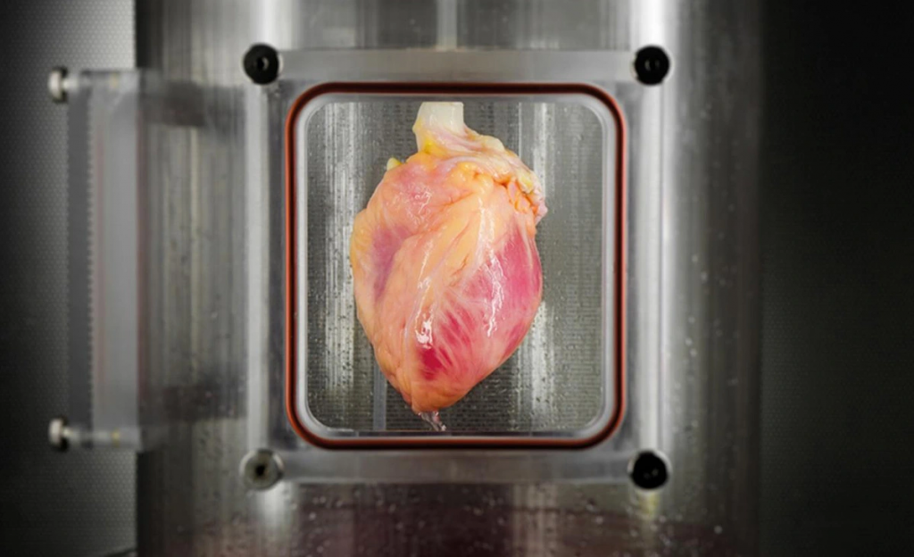
A heart derived from pluripotent stem cells being cultured in a bioreactor delivering a nutrient solution to replicate the environment a heart would need  (Bernhard Jank, MD, Ott Lab, Center for Regenerative Medicine, Massachusetts General Hospital)

Issues arisen from regenerative medicine

While current treatments and future turnouts derived from regenerative medicine are both incredible and life-changing, there are some set backs.

One of the main concerns is the safety of stem cell therapies, there is a risk of the cells differentiating into the wrong type of cell, which could lead to adverse effects such as tumours or other tissue damage. As the cells have been transplanted from outside the body, there is also a risk of infection or rejection of the transplanted cells by the immune system.

Another main concern is the ethics of stem cells taken from embryonic cells, due to the embryo needing to be created and destroyed for the extraction of the stem cells. However, this issue is currently being addressed by using alternative sources, with the use of induced pluripotent stem cells being tested currently.

Not only are there ethical and safety concerns but stem cells can also be very expensive. Due to the cost, it may limit access for some patients and be discriminatory against people who may not be able to afford the treatment.

Final thoughts

Throughout my research on this topic, I have formed an opinion that supports the use of stem cells and regenerative medicine. Although there are some cons of the treatments, I feel as though the pros outweigh them, as current treatments used today are saving lives, and with continued work from scientists and doctors worldwide, even more can be saved. Hassan would have been in constant pain without these treatments, but now he can live a long and happy life thanks to them. Not only can the benefits be seen through Hassan’s story, but also in the lectures in tissue engineering and stem cells by Dr.Evans.

“Stem cells are the future of medicine, but they are also the present. We have only just begun to scratch the surface of what these amazing cells can do, and the possibilities are truly limitless.” –

Dr. Robert Lanza

How can prosthetics be adapted for different lifestyles?

During a lecture and workshop focusing on prosthetics, I was intrigued by the design and functions of prosthetic limbs. Whilst watching the lecture, I spent some time wondering how the body communicates and works with the prosthetic to provide a function so similar to those of a natural limb in different everyday activities. Going into this research I didn’t have much prior knowledge and I perceived prosthetics to be limb replacements, but didn’t know their wider use. Upon seeing and interacting with a lower limb prosthetic in the workshop, I decided to further research this concept. 

Types of prosthetics

At the start of my search, I found there were many ways a prosthetic limb could work. Firstly, it can be powered by the body moving itself, e.g. where a cable may be placed on the shoulder and extend to a prosthetic hand. As the shoulder moves, the prosthetic moves. Secondly, it may have buttons, e.g. pressing a button on a prosthetic hand will cause the hand to grip an object. More recently, myoelectric powered prosthetics have been developed. This links muscles in pre-existing limbs to generate electrical signals and pulses via electrodes placed on the skin.

Example of myoelectric prosthesis.

Prosthetics for different lifestyles

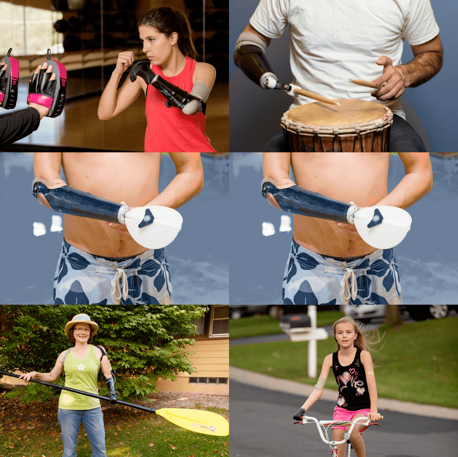
I then wondered how a prosthetic like these could be used in different scenarios and lifestyles, e.g with different hobbies. During my research I found the website Arm Dynamics which discusses the creation and execution of many prosthetic attachments for those with varied everyday lives.

Ways prosthetics have been adapted for different activities.

Being an active gym goer I wondered how prosthetics could be used efficiently at the gym to complete exercises with correct form and came across Max Okun. Max is a personal trainer who was born without a left arm and forearm, but living with this through his life wasn’t going to stop him in his passions. It did however cause him injury as he was overusing his right arm, to counteract this pain, instead of surgery, he decided to use exercise to build up his muscles. It was therefore important for the engineers creating his prosthetics to ensure whilst Max was doing the exercises he was not causing further injury. 

Max Okun Patient Profile from Arm Dynamics on Vimeo – This video shows Max using his prosthesis.

My reflections.

Researching this stream of engineering made me very grateful to be in a generation of such intelligent creators. As someone with fully functioning limbs, I think it is easy to take for granted how our brains are able to seamlessly communicate with our body parts. Even with tasks such as writing this blog, I require little to no thought in using my fingers. I can go bowling and tap dance without worrying about my mobility. I look forward to seeing what comes next in prosthetics and where it can go. Sitting this module has inspired me to look for careers that can aid in this development.

Spinal Motion in Post-Surgical Scoliosis

Scoliosis is an abnormal lateral curvature of the spine, causing misalignment. In order to be considered true scoliosis, the curvature, measured by the Cobb angle, must be more than 10 degrees. Spine specialists recommend different treatment options depending on this angle, with more severe and progressive cases resulting in surgery.

Examples of a mild, moderate, and severe case of scoliosis

Spinal Fusion

Spinal fusion, which has been the standard surgery for scoliosis for the past century, involves realigning and fusing the curved vertebrae with screws and rods. Although this procedure has good outcomes, with low complication and re-operation rates, it can diminish spinal mobility and flexibility. This means that those taking an interest in sports who undergo spinal fusion, for example, can lose their ability to perform. As someone with scoliosis myself (although mild), and having previously competed in tennis tournaments, this got me thinking about alternative spinal surgeries that do not have a negative impact on mobility.

An example of a patient before and after spinal fusion

The desire to maintain spine motion has fuelled the development of various growth modulation procedures. The goals of these procedures are to correct the spinal deformity and maintain motion.”

Scott. J. Luhmann, M.D.

Vertebral Body Tethering

As of recent, non-fusion spinal surgery has been used on those with scoliosis to correct curvature, while preserving flexibility and mobility. One method is through vertebral body tethering (VBT). This is where anchors (coated in hydroxyapatite, the substance that bones are composed of) are anteriorly attached to the vertebrae on the convex side of the scoliotic curve, with a flexible tether (made from polyethylene-terephthalate) connecting these anchors. The foundation of the procedure is that the tension from the tether slows the growth on the convex side of the spine, giving time for the concave side to catch up and driving the spine into the correct alignment. Due to the flexibility of the tether, and the absence of bone fusion, this allows for better spinal mobility post-surgery. Because VBT works via growth modulation, the most suitable candidates are those who have yet to reach skeletal maturity or are experiencing progressive scoliosis. This therefore means that VBT is less generalised than spinal fusion, and due to it being a new approach, there is some uncertainty with its long-term outcomes. Possible complications surround the fact that the tether may break through prolonged stress, though this does not pose much of an issue once spinal correction has completed. Early reports into the post-operative results of VBT do however demonstrate a high success rate and low revision rate.

Image showing what a scoliotic spine would look like before and after VBT
Video of the VBT procedure

Posterior Dynamic Correction and The ApiFix

Another non-fusion surgery for scoliosis is posterior dynamic correction, which occurs with the use of the ApiFix. The ApiFix is a self-adjusting rod, implanted on the concave side of the spine using a posterior approach, serving as an internal brace, and helping straighten the spine. The device can expand and so accommodates skeletal growth and additional correction. It is fastened to the spine with a single-level fusion (meaning only two vertebrae are being fused), with a total of only three screws needed. The rod features two polyaxial joints in which the vertebrae are fused, providing a greater degree of motion than spinal fusion. The ApiFix is most suitable for those with single spine curves and a Cobb angle between 35 and 60 degrees. Because this device and the procedure is novel, questions have arisen about its success rates. For example, possible complications include rod breakage and osteolysis (destruction of bone tissue). Early reports into the post-operative results of posterior dynamic correction with the ApiFix do however display its success and provides preventative measures to decrease failure and re-operation rates.

Image showing a scoliotic spine before and after posterior dynamic correction with the ApiFix
Video of the ApiFix procedure

Final Thoughts

As I researched into the topic of surgical treatments for scoliosis, I was pleased to find other options as well as spinal fusion that can help preserve the individuals spinal mobility and flexibility. I believe that it is important to offer various treatments, while considering patient suitability, in order to improve the patient’s condition while not having a negative impact on their lifestyle. With the nature of these procedures, it has given me insight into how individuals requiring scoliosis corrective surgery have to negotiate between greater spinal motion with some uncertainty in long-term outcomes, or a long-lasting solution with diminished spinal motion. Personally, I would decide to go with the former option, with spinal fusion becoming a last resort if long-term outcomes are not successful. Nevertheless, I am excited to observe the follow-up results of these newer procedures and the advances technology will have on scoliosis corrective surgery.