The University of Southampton

We all are chimaeras!

Karen Keegan

In 2002, a woman called Karen Keegan underwent a genetic test to find a suitable kidney donor within her family. Results were astonishing: Karen was not the mother of her sons.

Karen was later discovered to be a human chimaera with 2 distinct sets of DNA, with that in her blood cells different compared that of the other tissues.

The video below explains in more detail Karen’s story and introduces the phenomenon of chimerism

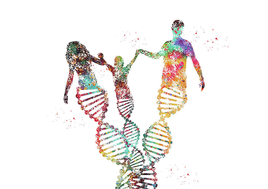
A chimaera is a single organism or a tissue which contains cells with at least two different sets of DNA. Since all animals develop from a single fertilised egg, every single cell of the body is supposed to have exactly the same DNA. 

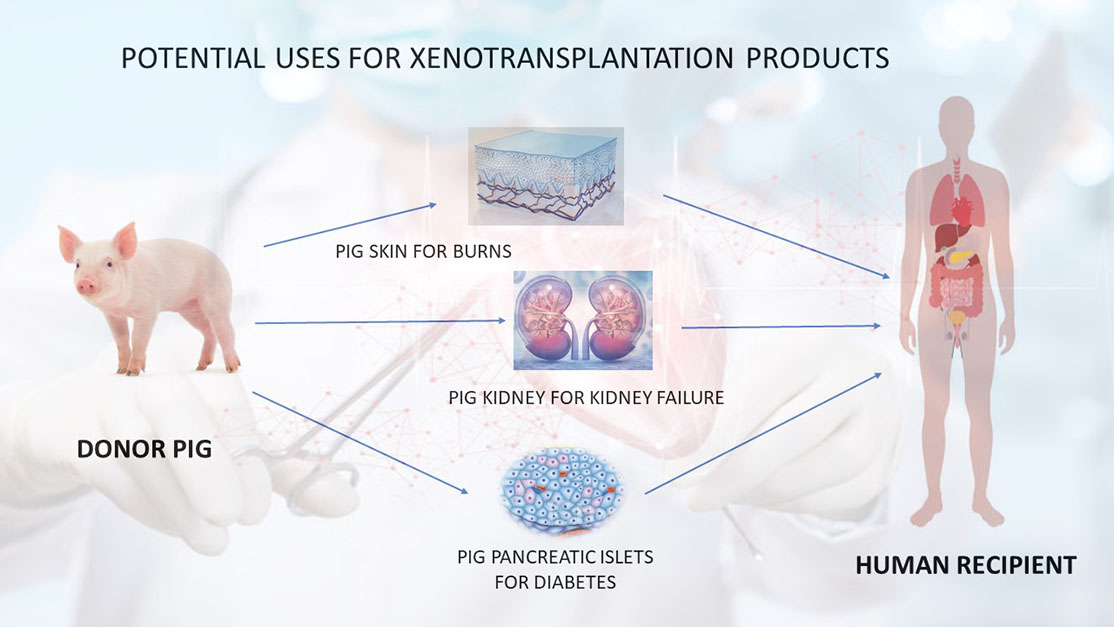
Researchers have developed chimaeras, whose bodies are a mix of cells from diverse species, in order to study human disease and find effective treatments. In fact, the most common organisms used to make chimaeras are mice, rats, pigs and monkeys, which all share a significant number of homologous genes with us. 

However, chimaeras are not always man-made, as there are at least three examples of already existing human chimaeras: 

  1. When a foetus absorbs its twin 
  2. When a patient undergoes a bone marrow transplant – e.g., to treat leukaemia  
  3. When a small fraction of cells are from someone else, so-called “Microchimerism”

1) When a foetus absorbs its twin

This occurs during a fraternal pregnancy when one embryo dies very early, and the remaining one “absorbs” some cells from its twin in the womb. Therefore, the remaining foetus has two sets of cells: its original and the one acquired from its twin. These individuals very often do not even realise they are a chimaera. 

Person with 2 different eye colours

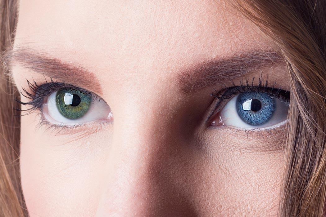
People with this form of chimerism very often do not even realise they are a chimaera because they can appear entirely normal. Most of the time, this is discovered by accident, and makes this particular phenomenon very difficult to quantify. More rarely, individuals show visible signs, such as diverse eye colours, patches of skin of different shades, or even a mixture of female and male cells which can cause abnormalities in their reproductive organs.

2) Bone marrow transplant

Human bone marrow contains haematopoietic stem cells, which give rise to all types of blood cells in our body (e.g., white and red blood cells). When a person undergoes a bone marrow transplant, their own bone is destroyed and replaced with a new one from another person, therefore acquiring “foreign” blood cells for the rest of their life. In fact, these haematopoietic stem cells are genetically identical to those of the donor but are different from the rest of the patient receiving the transplant!

3) Microchimerism

This phenomenon seems to occur in almost all pregnant women (at least temporarily), as a very tiny number of cells from the foetus migrate into her blood and travel to diverse organs. 

In 2015, researchers took samples on 26 women from their kidneys, livers, spleens, lungs, hearts, and brains. Results showed that all women, who had all been carrying sons, had foetal cells in all of these tissues, as these contained a Y chromosome (found only in males). 

Another study in 2012 found foetal cells in 63% of the brains of 59 women ages 32 to 101, including the oldest who was 94 years ago. This could suggest that most mothers have cells from their babies growing in various parts of their body after pregnancy, and these cells can survive in a lifetime.

Final considerations

Before hearing Karen’s story during a university lecture held a week ago, I had no idea about the existence of human chimaeras. Her story quite shocked me as I couldn’t believe that she was not really the mother of her son, and therefore I decided to dig down the topic of chimaerism. 

In the past, I had heard the term “chimaera” in some fantasy movies, as I am very passionate about this genre. However, I had never thought that this was possibly achievable in the real world, not even artificial chimaeras made in the laboratory. 

Through this research, I learned that Karen’s story is not the only one of human chimaeras but there are many other instances. Thinking about what to write in the final considerations for this post, I had an epiphany, which inspired the title of this post: at least temporarily, we all are chimaeras! 

We exchange living cells between us more often than we think, ranging from kissing or touching to sexual intercourse or a more invasive transplant! Yes, you read it right, but you probably didn’t know that even a very passionate kiss is able to transfer living cells from you to your partner and vice versa, making both of you temporary chimaeras! So I’ll be very careful the next time I choose who I’ll kiss … 

Now, I recognise that chimaerism can come in various forms and quantities. More often, we contain only a few living cells from another person, and more rarely a single person can be a fairly equal mix of cells with distinct sets of DNA. 

Building the first layer of knowledge on a completely new topic has completely challenged my previous understanding. I learned that hearing something for the first time can be quite shocking at first but I should make use of this experience positively to expand my views and opinions, rather than as a way to discourage myself. In the future, I will keep this positive attitude to get to know new topics, which could completely challenge the knowledge I have just acquired!

References:

1. Scientific American article written by Rachael Rettner, LiveScience on August 8, 2016

https://www.scientificamerican.com/article/3-human-chimeras-that-already-exist/

2. MCSM RAMPAGE article written by Fariha Fawziah on January 31, 2017

Healing Blindness with a 3D cornea

The scientists’ 3D printed cornea transplant. Photo via the Times of India.

From the lecture on tissue engineering, I found impressive the number of applications of 3D printing: I had no idea that this technology could be used to create entire human organs for transplantation!  This led me to look more into recent advances in 3D printing, where I came across this article1 that described the first 3D printed cornea developed in India. 

How was this 3D cornea developed?

In the video below (edited from this YouTube video), I explain the development and wider consequences of this 3D cornea. 

While writing the script and editing the above video, I started to wonder how regulations about consent to donate human stem cells for research purposes differ from those described in the bioethical lecture, including the Human Tissue Authority instituted in 2005 in the UK.

According to this government’s website2, guidelines for stem cell research in India have been established by the Indian Council of Medical Research (ICMR) and the Department of Biotechnology (DBT) since 2007.

Both in the UK and Indian regulations, the donor must be aware of the specific research purposes and is required to be mentally capable of giving consent. This made me realise that stem cells are worldwide considered very powerful tools for scientific advancements but their use must be tightly regulated by multiple laws and authorities to prevent unethical research.

A Cornea Stromal Lenticule 3D printed by Indian firm Pandorum Technologies.

While reading this journal’s article3 to dig down more on how the cornea was made, I found out that the newly developed bio-ink can be used as a new treatment. It is able to save the patient’s sight by sealing the corneal perforation to prevent infection during war-related injuries or in a rural area without nearby eye care facilities. I had never thought that materials or tools created to address a specific research aim could become discoveries for a new patent themselves!

What are the advantages & disadvantages of this 3D printed cornea?

Since multiple news articles1,3,4 celebrated the development of this cornea, I was curious to know whether this technology could address all the concerns coming out of my mind: Is it safe? Is it biocompatible? Is it comfortable? Therefore, I decided to search for pros and cons using two different sources: a more subjective journal’s article3 versus a more objective scientific paper5

Advantages3Disadvantages5
1) Natural, biocompatible, free of animal residues, and safe
2) Printable in different diameters (from 3mm to 13mm) to meet diverse requirements
3) Each human donor stem cell can contribute for the preparation of three 3D printed corneas
4) The tissue coming from donor corneas needs to meet lower optical standards compared to those required for a transplantation
1) Uncomfortable to wear
2) Transplantation process is complex
3) Multiple surgeries and long-term topical medication is often required
4) Limited field of view
5) Unsatisfactory aesthetic appearance
6) Potential for post-operative complications such as extrusion and glaucoma
Protesters in India against animal tested products

From this insightful learning journey, I realised that a 3D printed cornea could become a concrete solution in future years to bypass the difficulties of finding a donor match and the complications associated with synthetic corneas. For instance, a good surgeon should correctly perform transplantation surgery, and the unaesthetic appearance or the limited field seems to be better than the alternative of blindness. However, the side effects of medication and post-operative complications could significantly delay a potential application in humans any time soon. 

All news articles1,3,4 emphasise that one of the most important benefits of the 3D cornea is that it is free of animal residues. I  did not know that animal products cannot be used for research purposes in India, since this is perfectly accepted in the country I live in, the UK. I learned that there are many other factors that can significantly affect scientific research, including religion or social acceptability. As a future biomedical scientist, I will keep this in mind while designing new products, tools or drugs in order to respect the target countries’ policy, religion, culture and tradition.

References

  1. 3D Printing Industry: “Scientists say India’s first 3D printed cornea can be used in humans after successful animal trial”, written by PAUL HANAPHY, on August 16, 2022. More information at this link: https://3dprintingindustry.com/news/scientists-say-indias-first-3d-printed-cornea-can-be-used-in-humans-after-successful-animal-trial-213745/
  2. Association for the Advancement of Blood & Biotherapies. More information at this link: https://www.aabb.org/regulatory-and-advocacy/regulatory-affairs/regulatory-for-cellular-therapies/international-competent-authorities/india
  3. tct magazine: “3D printed human cornea developed by team of clinicians and scientists in India”, written by OLIVER JOHNSON on August 17, 2022. More information at this link: https://www.tctmagazine.com/additive-manufacturing-3d-printing-news/latest-additive-manufacturing-3d-printing-news/3d-printed-human-cornea-developed-by-team-of-clinicians-and-scientists/
  4. The Times of India: “Hyderabad scientists develop India’s first 3D-printed cornea”, written by Syed Akbar on Aug 14, 2022. More information at this link: https://timesofindia.indiatimes.com/city/hyderabad/hyderabad-scientists-develop-indias-first-3d-printed-cornea/articleshow/93560273.cms
  5. Holland, Gráinne & Pandit, Abhay & Sánchez-Abella, Laura & Haiek, Andrea & Loinaz, Iraida & Dupin, Damien & Gonzalez, Maria & Larra, Eva & Bidaguren, Aritz & Lagali, Neil & Moloney, Elizabeth & Ritter, Thomas. (2021). Artificial Cornea: Past, Current, and Future Directions. Frontiers in Medicine. 8. 770780. 10.3389/fmed.2021.770780.

Pig’s kidney in humans?

“It just looked like any transplant I’ve ever done from a living donor. A lot of kidneys from deceased people don’t work right away, and take days or weeks to start. This worked immediately.”

Dr. Montgomery, director of the N.Y.U. Langone Transplant Institute in Manhattan. 

Currently, more than 100,000 Americans are on the waiting list for a transplant, with more than 90,000 only needing a kidney. 12 people on this waiting list die each day. 

Researchers have been studying to grow human organs in pigs to create perfectly suitable tools for transplantation into humans. So far, pig hearts and kidneys have been successfully transplanted into baboons and monkeys but never in humans.

In 2021, an incredible surgery was performed in N.Y.U. Langone Health. The aim was to “simulate” an actual transplant procedure to prove that a genetically engineered pig’s kidney is able to avoid rejection by a human’s body. This specific kidney was obtained by knocking out a gene that encodes a sugar able to elicit an aggressive human rejection response. This was done because rejection occurs very often in the so-called “xenotransplants”: transplants from animals like pigs and primates to human beings.

A surgical team at the hospital in New York examined a pig kidney attached to the body of a brain-dead recipient for any signs of rejection.

A pig’s kidney was attached to blood vessels in a brain-dead patient’s upper leg, outside the abdomen. Then, the surgeons covered it with a protective shield in order to allow constant observation. Kidney’s function was measured by taking samples over the following 54 hours study period. It was the first operation of this kind ever in human history.

Results are astonishing: the kidney was working normally “almost immediately” by starting to make urine and the waste product creatinine. Importantly, there were no signs of rejection.

Video showing the successful transplant on local news 

Reactions among the transplantation experts

According to the experts, this is a very good sign as an organ functioning outside the body is a strong indication that it will work inside the body. While some surgeons speculated that this practice will become ordinary in the next few years, others argue that still much more work is needed. Anyway, all transplantation experts acknowledged the importance of this mind blowing surgery. Among them, reactions ranged from extremely optimistic to very careful and warning.

“This is really cutting-edge translational surgery and transplantation that is on the brink of being able to do it in living human beings”

Dr. Amy Friedman, former transplant surgeon and chief medical officer of LiveOnNY

However, this looks too good to be true …

Testing experimental kidneys in humans has presented several daunting concerns:

  1. We still do not have studies and answers about long-term consequences of such an operation 
  2. Animal welfare and exploitation, as thousands of pigs will be raised with the unique aim of getting their organs harvested for a “better” human receiver 
  3. Pigs can get affected by some viruses which do not affect humans. Can then humans develop these viruses or are they protected? 
  4. Last but not least, we need to make sure that the person fully consents to the practice, knowing all the benefits and risks. In this specific experiment, the family gave the consent and the person themselves, as they were brain-dead. How could this work in practice in the future?

This opens the door for using genetically engineered pigs as a sustainable renewable source of organs for severely ill patients, as the current clinical need is still unmet. However, this procedure will not be available to any patient any time soon due to many relevant medical and regulatory hurdles that still need to be overcome.

In the future, not only kidneys, but also other organs could be transplanted, including hearts, lungs, livers … potentially every organ in our body also present in the pig! All these organs could save the life of thousands of people who die everyday waiting for a suitable human donor, as there is scarcity of human organs. For instance, most dialysis patients do not qualify for transplants, which are reserved for those more likely to survive after the procedure.

Final considerations

At the end of a university lecture, the professor briefly quoted a recent research which tried to create a complete human organ into a pig by combining human iPSCs (Induced Pluripotent Stem Cells) with a pig embryo. The experiment actually failed, as there were pig cells stained in the human-like organ. However, this sparked my curiosity to find out more about xenotransplantation between pigs and humans to see if there have been more recent successful experiments. 

One shocking aspect I discovered while writing this post is that an organ is able to properly function without even being “internally” transplanted but simply being linked with blood vessels to the natural functioning organ. I had never thought this was possible, nor had I never read about  a similar experience. 

My expectation was to discover that someone was at least being transplanted with a very small and simple organ from a pig. Although the surgery is not an actual “transplantation”, it clearly represents a sea change for the organ transplant’s knowledge. Through this research, I have challenged my previous viewpoint about scientific research because I learned that progress sometimes requires “intermediate” steps to achieve the final goal. In the future, I will positively approach all research and experiments, although these don’t necessarily meet my initial expectations.  

What about you, would you ever receive a organ from a pig, or a primate?

Reference

“The New York Times”, article written by Roni Caryn Rabin on October 19, 2021

Can prosthetics learn over time?

An ongoing experiment

The “Esper Hand”

“We sought to create a light and durable hand with human-like dexterity that learns over time and can help people with limb differences live their best lives confidently.”

Dima Gazda, co-founder of Esper Bionics

In 2021, New York-based engineering startup Esper Bionics developed a new incredible bionic hand that seems to work three times faster than many of all its other competitors currently available on the market. 

In the video below, a user called Nika performs a series of tasks with her bionic Esper Hand, ranging from grabbing small objects to wielding a beefy knife.

Esper Hand has 5 flexible and modular fingers and weighs only 380 grams, making it lighter than a human hand and many limb prosthetics on the market. It is composed of a combination of aluminium, fluoroplastics, polyoxymethylene plastic, titan, nylon, bronze, steel and 3 different types of silicone. On top of interfacing seamlessly with its wearer, Esper hand has also 4 different sizes and 5 colours.

This hand can rotate and grip in several ways and the modular fingers can form multiple common grasps including cupping, flexing, making a fist and pinching fingers together. This allows the wearer to perform a wide range of movements for day-to-day tasks, including using kitchen utensils, opening bottles, tapping a phone screen or even driving a car!

This prosthetic can also be easily disconnected via a further mechanism so that the wearer can switch it on and off according to their needs, such as while changing clothes. 

How does “Esper Hand” work?

Electromyography-based brain-computer interface (BCI)

This bionic hand was developed by conducting durability tests on 3D-printed versions of the hand to adjust its size and shape. Esper Hand makes use of an electromyography-based brain-computer interface (BCI), a computer-based technology system that collects brain information or activity, in order to trigger muscle contraction. This type of technology is not completely new, as it has already been used by paralysis patients to control machines by simply their thoughts.

When the wearer wants to control their hand, the brain sends electrical signals to specific muscles in order to activate them. Esper hand is able to simulate this brain control through  over 30 non-invasive myoelectric sensors that connect the stump socket to the wearer’s skin. These are the myoelectric inputs for movement which generate electrical signals exactly as those naturally generated by one’s muscles to trigger action in the hand. 

What makes “Esper Hand” so special?

“Thanks to all the collected data, the platform updates the control algorithms of the hand so that next time the preferred grip will have a higher priority to be chosen in the same situation”

Dima Gazda, co-founder of Esper Bionics

This prosthetic works with intuitive self-learning technology that can predict intended movement faster than similar prosthetics, therefore improving its performance over time on the same user. 

This occurs via a specific phone app that sends all the myoelectric inputs to Dubbed Esper Platform, a cloud based-platform managed directly by Esper Bionics, which collects and stores data about the user’s movements. This allows its creators to study and improve the algorithms that translate those inputs into digit movements so that the wearer’s next action will be quicker predicted and performed, basically like a “learning” mechanism. 

Personal reflection

After attending a university workshop where the professor showed us examples of prosthetics, I felt quite fascinated by the unrealistic appearance of a prosthetic hand that was used by a double amputee to cut a carrot. A example of this hand is shown in the picture on the right. It is very common in US rural areas because it is extremely functional. However, it is not very aesthetic in appearance since it looks more like a very old hook than a beautiful human hand.

The same day of the workshop, the video about Nika using Esper Hand appeared on top of my YouTube page. That girl seemed so happy and comfortable using Esper hand (which really resembles a human hand!) that I chose to dig down the topic.

During the research, I felt quite shocked by knowing the possibility of a way to improve a prosthetic’s performance via more efficient algorithms. I had never thought about it before, and I realised that this is a very clever and non-invasive method not only to improve the prosthetic, but also to learn more about research of human movements in order to increase scientific output!   

This realisation inspired the title of this post: “Can prosthetics learn over time?”. Incredibly, the simple answer was: “Yes!”. However, this was not enough for me, and I started to wonder about humans made by prosthetics that I had seen in Marvel movies, such as incredible superheroes fighting against the most powerful devils in the world to eventually restore peace.

What if this bionic hand could actually perform even BETTER than the natural hand? 

I thought that if prosthetics not only could really improve their performance over time to equate that of the natural hand, but also overcome the performance of the natural hand thanks to outstandingly efficient algorithms… This might open the doors for a new technological era in the AI and robotics field. 

However, computers and AIs are much quicker at performing logical calculations and computations but not interpreting signals from the outside world to generate an adequate response, as our brain does continuously. Some studies have actually proved that the brain has higher computational power efficiency than electronic computers by orders of magnitude. This is why computers and AI architectures are all modelled in an attempt to better emulate the human brain! 

Getting to know the Esper hand story has completely challenged my previous understanding of the potential of prosthetics. I learned that prosthetics are not static entities but can “learn” over time, although in a completely different way than a human brain. This triggered me so many unsolvable questions and made me wonder in my fantasy. In the future, I will keep this curious attitude by letting my thoughts wander for a while to come up with interesting observations that can lead me to further research and understanding, which is not necessarily related to the initial topic. 

Anyway, I will also need to be careful when I should stop my fantasy to be more realistic in order to avoid an unproductive combination between fantasy and reality. For example, the presence of “superhumans” looks still very far from reality, as many of our brain’s processes are still unknown. This strongly affects our knowledge of even the most powerful and quickest prosthetic AI. But in the future, who knows, maybe the “superman” from the comics will exist for real!

References

Dezeen article, written by Alice Finney, on February 24, 2022

https://www.dezeen.com/2022/02/24/esper-bionics-human-prosthetic-arm-mind-control/#:~:text=Esper%20Hand%20is%20a%20prosthetic%20arm%20that%20can%20be%20controlled,an%20action%20in%20the%20hand.

If you are interested to know about the “Esper Bionics” company, you can find more information in the following link: https://esperbionics.com/.

Blade Runner

Oscar Pistorius, the “Blade Runner”, is a South African runner who had both his feet amputated when he was 11 months old due to a congenital defect. Since then, he started to run in sprints events for both below-knee amputees and non-disabled.

Since Pistorius was starting to win over the best able-bodied athletes, people have started questioning more about the role of prosthetics in athletics. In the media, there was an important controversy whether prosthetics give unfair advantages to amputees over able-bodied athletes. 

Can really runners with prosthetics obtain a better performance than able-bodied athletes?

After Pistorius was banned from competitions in 2008, scientific research was conducted to discover if his prosthetics were really giving him any advantages. 

A research group led by the biomechanics Alena Grabowski, where Pistorious himself took part in, aimed at comparing his abilities to those of some able-bodied track athletes. Three distinct tests were performed to measure a series of parameters: 

  1. Energy cost in running → measured by breathing and metabolic of runners during short sprints;  
  2. Endurance → measured by setting a treadmill on the maximal speed that runners could maintain; 
  3. General running mechanics → observed by asking runners to augment their speed on a treadmill until they could no longer take eight consecutive strides without maintaining their position.

Results of this study proved that Pistorius’ running capabilities are not significantly different from those of able-bodied runners. This allowed him to end his ban and come back to compete.

London 2012 Olympic Games: 400m race

Pistorius was the first amputee to win a non-disabled world track medal. This happened at the 2011 World Championships in Athletics. 

Oscar Pistorius became the 1st double amputee to compete in the Olympic Games, specifically the 400-metre race. 

… Research on prosthetics is not over 

After this initial research, Grabowski conducted another study to test the influence of some parameters in a prosthetic on a runner’s performance. First of all, she modelled the foot as a spring system in order to pick the crucial parameters of a prosthetic to modify: speed, stiffness, and height.


The research consisted of testing five runners on a treadmill, increasing the speed on each trial until they could no longer hold their position. This same experiment was repeated by changing the different parameters of the prosthetics until enough data was collected to compare. 

Findings of this study are very interesting: 

  1. The length of the prosthetic did not have any effect on running speed;
  2. The stiffness seemed to help runners only at medium/low running speed but not high running speeds.

Therefore, a prosthetic gives a significant advantage for long distance runners but not for short springs. 

Final considerations

I chose the module “Engineering Replacement Body Parts” because I have always been fascinated by the world of prosthetics. After the lecture focussed on hip and knee implantation and leg prosthetics, I realised that there are so many requirements that must be addressed during the design in order to make a functional prosthetic. However, the most important factor that engineers consider is always: “What does the patient want to achieve?”. 

The world of prosthetics can open up many different doors for amputees: from addressing simple day-to-day tasks to competing alongside able-bodied athletes in the Olympic Games. Before researching more in detail about Pistorius’ stories, I had already heard his name multiple times but I was probably too young to remember his participation in the Olympic Games in 2012. As I saw this familiar name in my research results, I decided to dig down more about this very fascinating topic. 

This research was extremely illuminating because I learned from scratch a completely new world under several aspects. First of all, I had no idea that amputees could compete with able-bodied athletes or at least not in such prestigious competitions like the Olympic Games. Secondly, I was shocked that an amputee actually competed with able-bodied athletes because I had never heard or seen it before. Thirdly, I initially believed that prosthetics didn’t give any advantages to the amputees but only disadvantages over able-bodied athletes in terms of running performance! 

Grabowski experiments’ results were the most shocking part for me but also the most intellectually challenging. I learned that scepticism on athletes with prosthetics competing with able-bodied athletes at the Olympics has not been scientifically proven, as the effects are minimal or even non-existent in the case of sprinters. I believe that this is the reason why experiments conducted by Grabowski and her team are so important. Moreover, this research was conducted only because Pistorius was a champion who wanted to fight for his rights to compete in such a prestigious competition like the Olympics. However, very likely there are many more other athletes over the world, and none of this has come to light as much as Pistorius.  

I believe that regulations of all competitions should follow scientific facts and research over emotions and fear. Amputee athletes should be allowed to compete with able-bodied athletes until there is concrete evidence which proves the opposite. In the future, I will keep the rational attitude I used while doing this research to write this post in order to effectively welcome and learn from any piece of scientific research that completely reverses my initial thoughts!  

In the following years, with further development of prosthetics, I am sure there will be much more research about prosthetics in sport, as more amputee athletes will be able to reach the same level of performance as able-bodied athletes. So far, an amputee athlete has never won over able-bodied athletes at the Olympic Games, not even a champion like Pistorius. With the help of scientific research … Who knows, maybe some amputee athletes could change the Olympics history in the future!

Reference

University of Nottingham, page: “Biomechanicals in the Wild”, article written by Kaleb Schoolman, 2019

The Resilience Project

Doug Whitney

“Doug Whitney has a terrible family legacy. Many of his relatives carry a gene mutation that causes early-onset Alzheimer’s disease, a severe form of Alzheimer’s that strikes in one’s 40s and 50s. The disease has wreaked havoc on his family, killing his mother, older brother and more than a dozen of his aunts, uncles and cousins. In 2011, Whitney learned that he also carried the mutation. But at 67 years old—years past when family members began to show symptoms—Whitney’s memory is intact.”

The Resilience Project

How is Whitney’s story possible?

Some people have genetic changes that protect them from getting specific diseases or more able than most to recover from challenges to their health. 

These people are called “RESILIENT HUMANS” 

The “Resilience Project” aims to discover why some people are more able than others to resist or recover from challenges to their health and escape disease.

Finding and studying resilient individuals could pave the way to new insights about health, better disease prevention strategies and new treatments. This is why scientists hope that studying their resilience will inspire new treatments for diseases. 

Factors affecting a person’s resilience … much more than genetics and biology!

Research shows that our health and our resilience to disease are influenced by a number of factors:

  1. Social circumstances: people with stronger social connections tend to live longer, are more likely to survive a heart attack and are less likely to suffer a recurrence of cancer.
  2. Environment: access to healthy food, green spaces and gyms can boost resilience to disease –  e.g., reduce the risk of diabetes
  3. Medical Care: access to therapies and drugs 

I discovered the existence of “The Resilient Project” around one year ago because it  was briefly quoted during a lecture. This has been stuck in my mind since then, as I immediately felt quite shocked getting introduced to some of these incredible humans’ stories, such as Whitney’s experience. This strong curiosity made me read other resilient stories and inspired me to research more about the “The Resilient Project”.  

Further research on The Resilient Project’s website completely challenged my previous assumptions about human disease. I believed that only genetic factors could significantly contribute to the development of a disease, together with minor contributions from the environment as the initial trigger. I also had never thought about medical care as a relevant component of a disease. However, this can be life-saving in specific conditions of isolation such as rural areas, or even third world countries with an insufficient number of clinics and hospitals and lack of appropriate medical equipment.

More incredibly, I realised that there are so many other factors that can affect our resilience but on which we can exercise total power, such as social circumstances and individual behaviour. For instance, we can choose our circle of relationships, from our partner and best friend to the furthest acquaintance. Amazingly, this can not only extend our lifespan but also protect us from developing very severe and common diseases, including cancer and heart attacks!  

Through this research, I learned that human diseases are extremely complex and cannot be caused by a single factor, such as our genes. Having challenged this assumption will help me to stay open-minded while examining scientific papers. As a future scientist, I will keep in consideration that many other variables are involved in diseases, and only a small percentage is under my control, such as developing new drugs based on pure genetics and observable biological mechanisms. 

In conclusion, I believe that writing this post to openly talk about “The Resilient Project” can increase people’s awareness about the existence of such unthinkable humans. Everyone can potentially know another resilient human or be a resilient human themselves! This could tremendously help current research by simply contacting this association in order to make concrete outcomes for other people across the world who are suffering from the same disease.
If you are interested to know more about the “Resilience Project” or read stories from resilient humans, you can find more information in the following link: https://www.resilienceproject.com/resiliencestories/.

It’s a knockout!

The human genome is “packed” in a particular molecule called DNA, which composes the core of our cells. This molecule is extremely big; if we could completely stretch it, it would be 2 metres long! 

This incredible molecule, DNA, is composed of 2 main regions: coding and non-coding regions. The non-coding region is significantly more abundant and composes 98% of the total of our genome, while the coding genome only the remaining 2%. However, the coding region is hugely important, as it makes proteins, the basic functional units responsible for the smooth running of our body.

How do we identify and study the function of each coding region of DNA that makes a specific protein?

The scientific method utilised is called “knockout”, which refers to the use of genetic engineering to inactivate or remove one or more specific genes from an organism. In order to explore the function of a particular gene and study the effect of its loss, scientists historically used genetic engineering to create knockout organisms to study the impact of removing a gene from an organism, allowing the discovery of a gene’s functions.

For instance, rather than study the gene directly, it is easier for scientists to study the final product of a gene, its protein. Moreover, since the cell is composed of thousands of different genes that encode a massive range of proteins, how can we identify the function of THAT specific gene without concretely isolating it from the such chaotic “crowd”? 

The most commonly used animal model for knockouts is mouse as, incredibly and totally unexpected, it shares approximately 70% of the same protein-coding gene sequences with us. 

How do we make a knockout mouse?

Although knockout mice are very useful tools, they still have their disadvantages:

  1. Genetic engineering is a difficult, expensive and time-consuming
  2. Unethical issues due to the use of animals for scientific research 
  3. Although the degree of gene homology is enormous between mice and humans, there are still genes that could have a certain function in laboratory animals but not the same function in humans

Absent or non functional proteins are the cause of more than half of human’s diseases. This is why knockout animals are so important for biomedical research. Once the function of a gene is known, researchers can work on developing a range of drugs to treat the disease, such as drugs that replicate the function of the missing protein or drugs that mitigate the effects of the disease.

Conclusion

Last year during university lectures, I had heard for the first time the term “knockout” without having a clue of what it was. In the first lecture of this new module, Engineering Replacement Body Parts, I was explicitly introduced to this concept for the first time. 

My interest to research and expand knowledge on this methodology was that I had found this concept completely counterintuitive, as it works in a “reversed” manner! The principle is that we cannot study directly what the gene encodes if it is functional but we can infer its function when its final product, the protein, is no longer functional or completely absent. 

Initially, I thought that scientists were always able to “isolate” the molecule of interest and then test it under a range of conditions in order to discover their properties and functions. This assumption was harshly challenged by the fact that, unfortunately, isolation of molecules is rarely achievable for a series of obstacles, such as lack of powerful enough technology or extremely high costs. For instance, we cannot visualise “in real time” the action of very tiny cellular components, such as nucleic acids, but we can do it for larger entities like living bacteria under a microscope. 

I realised that something, which could immediately appear as counterintuitive, is not necessarily extremely complicated to understand. In the future, I will not feel discouraged if I cannot instantly catch a counterintuitive principle or technique, as this could reveal it as extremely important! Deepening my knowledge of knockout revolutionised my understanding of an essential scientific methodology used worldwide to discover the cause of many genetic diseases. As a future biomedical scientist, I will apply this knowledge to real-world scenarios not only to perfectly understand and review others’ research but also to design my future experiments and discover the cause of some human diseases!

Reference

National Human Genome Research Institute: https://www.genome.gov/genetics-glossary/Knockout

Engineering Replacement Body Parts

As a second year psychology student, I was provided many choices for my semester 2 optional module. However, I became torn between cognitive neuroscience and engineering replacement body parts. I ended up choosing this module as I wanted to learn more about prothesis and stem cells. Recently, I have been watching many episodes on NHK wherein prothesis was being discussed and I found these very interesting, ultimately influencing my decision to choose this module. One of the episodes focused on a guitarist who was born with congenital limb deficiency yet she is able to play guitar with a device that her dad developed. As someone who is also interested in guitar myself, I found myself engaged in the episode and wanted to learn more about prothesis and other aspects that compromise engineering replacement body parts.

CBT – Retraining the Brain

Cognitive Behavioural Therapy (CBT) is a well known form talking therapy, utilised for the management of a range of problems, by changing the way you think and behave.

Video Explaining CBT

There are a number of disorders that can benefit hugely from CBT, including OCD, phobias, eating disorders, panic disorders and depression. As somebody who has undertaken and subsequently completed their CBT journey, I will forever be an unwavering advocate for this form of therapy. Not only do you benefit from having a safe space to talk to a trained professional without fear of judgement, you learn new habits and methods that allow you to overcome your problem for the long-term. Completing CBT will leave you with a retrained brain, one that uses healthy habits and coping skills, instead of reverting to the dangerous behaviours that may have previously derailed your life.

Does parenting style affect brain development in children?

Brain development in early life has always fascinated me. It’s intriguing to consider the extent that environmental factors, in particular social interaction, dictate neurological development. Is it nature or nurture? The first episode of the Netflix series, Babies (2020), titled ‘Love’ delves into the biology of a mother and father’s love for their baby. It touches on the work of the GUSTO project, which comprised of a series of social experiments investigating a mother’s interactions with her baby. It also analyses different parenting styles, specifically their attentiveness to the babies. It shows some mothers constantly checking their babies, and others leaving them to their own devices. The less attentive babies were visibly calmer and quieter, which I found interesting despite unsettling. Along with a series of MRIs on the babies brains, the experiment concluded that the less attended to babies had larger hippocampi, the area of the brain which deals with stress. This is theorised to be because they have to deal with discomfort and stress by themselves, and due to the plasticity of developing neural circuits, the brain has tried to accommodate this. I found this study deeply fascinating, and as an adoptee, it forced me to consider my own experience in an orphanage and how it may have altered by own physiology at such a young age.