The University of Southampton

Engineering Replacement body parts 2024-2025

An interdisciplinary module

Bioengineering with Bubbles!

After an insightful trip to the hospital for the Stem Cell and Regenerative Medicine Masterclass, I discovered a new branch of biomedical engineering that I’d never considered: using bubbles in medicine! Now I know what you’re thinking, but these aren’t bubbles that you’d make from soapy water and by waving a wand around vigorously in the air. Instead, these bubbles are microscopic, made from a monolayer of phospholipids that self aggregate to form a sphere with a gas core. PhD student Aya Ben Issa demonstrated how to create these bubbles, alongside explaining some potential uses, including her own research focus, using them to break up biofilms. This got me thinking, what other uses for microbubbles are there?

Microbubbles for Imaging

Primarily, microbubbles are used for contrast enhanced ultrasound in medical imaging. Sounds like a mouthful, but in reality, it essentially improves the imaging quality by creating a better distinction between the tissues and the injected microbubbles. I’ve included a video so you can have a look for yourselves:

Microbubbles for drug delivery

When the idea of microbubbles was first introduced, my mind initially went to using them for delivering drugs. And why not? These micro compartments seemed like the ideal packaging for doing exactly that. With further research into the topic, I discovered that I wasn’t the only one who thought the same. Our current module lead, Professor Nicholas Evans has published an article on using them to deliver oxygen to tissues. This has shown promise for bone cancer treatments by reducing metastasis. This supports my initial thought; that things can be delivered within the bubble. But what about on them? Well, once again, studies have indicated that insertion of drugs into the microbubble shell in addition to attaching site-specific ligands have been show to allow targeted delivery of drugs to areas where pathologies have occurred.

When do Ultrasound and Microbubbles Go Hand in Hand?

So, you’ve put your drugs within your microbubbles and injected them into the patient. They’ve travelled to the target site. Now what? You need to actually get your drugs out of the bubble and into the tissue. This is where ultrasound comes in. Ultrasound is able to create oscillations and compressions within the shell of the microbubble, causing a large dose of drug to be released. In addition to the microtubules being the delivery agents of the drugs, they can also be used as mechanical agents. The ultrasound once again causes oscillations, this causes transient, miniscule disruptions in the plasma membranes of the cell, allowing drugs to once again enter the cell. This process is known as sonoporation.

Examples of Uses

In summary of the topics I have suggested above, the image below highlights some of these applications, from drug delivery to sonoporation, as well as some other potential applications.

What do I think?

In my opinion, the application of microbubbles have great potential. Of course, further studies will have to be done to fully understand the extent to which we can use this technology. There are still limitations when it comes to the therapeutic uses of these microbubbles in addition to some novel ideas such as Aya’s application of them to disrupting biofilms. What do you think? Can you think of any other uses of these microbubbles?

Want some more info?

Use the hyperlinks in this article for a more in-depth understanding of the topic I have discussed today! 🙂

A Reflection on Human Head Transplants

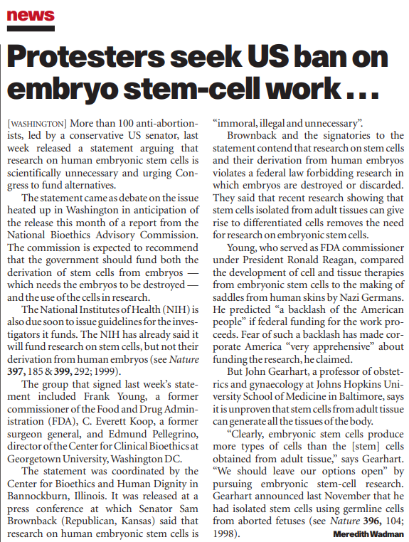
Following discussion on organ transplants, my reading led me to the debate of head transplantation, which, to those with terminal disease but a healthy head and brain [1], has the potential to extend life. It would be a last-resort treatment for patients whose body is affected but whose mind and head are healthy, hence treating conditions such as quadriplegia, progressive diseases, and inoperable cancers that have not extended to the brain [2]. In 2019, Valery Spiridonov—a man with Werdnig-Hoffmann disease, a muscle-wasting condition—pulled out of what would have been the first attempted human head transplant [3].

The surgery would require an immunologically matched brain-dead donor with a healthy body, onto which the head of the patient can be transplanted. The procedure has been performed on animals without long-term success [4], and in 2017, it was completed on a human cadaver [5]. Gkasdaris et al. discussed human head transplantation and highlighted the surgical issues involved as donor body selection, head and body interventions on the recipient and donor, ischemia time (managing the blood flow between the head and donor body), spinal fusion and spinal cord reattachment, and post-operative issues [1]. Beyond the surgical hurdles, ethical concerns are raised; notably, the EANS ethico-legal committee concluded that it is ethically unacceptable to attempt a head transplant [6]. Literature considers the great risk involved during and after surgery and the reality of living with the body of another person; for example, a 2017 study by Wolpe considers the extent to which our body, alongside the brain, “makes us who we are” [3]. Legality must also be taken into account. Wolpe points out that the surgery involves intentional decapitation, which means that, by definition, the death of a patient would be murder.

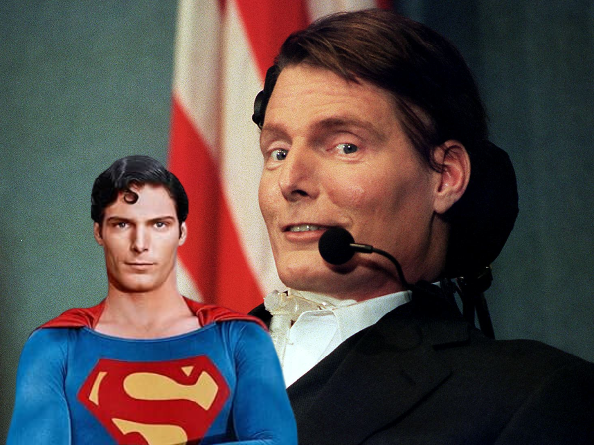
The idea of a head transplant is one that we, as the general public, expect to see in science fiction media, and would therefore view as something detached from reality. Frankenstein’s monster is often referenced in this discussion. We would consider the consequences of such a narrative—written to shock and scare us—and conclude that this is not something that should happen in ‘real life’. We have been told by the fiction we grow up around to be wary of the scientific unknown and the ‘mad scientist’ archetype who would attempt something so shocking and seemingly without consideration of ethical questions. It is difficult to suspend these preconceived ideas of what should and should not be done, and dull the instinctive, fearful dismissal of an attempt. Just the phrase ‘head transplant’ elicits shock, even as somebody so used to hearing about extreme surgical procedures. Through reading and consideration, I hope to form my own opinion on the subject, beyond general concern and morbid fascination.

I believe that the surgical challenges are the lesser issue when it comes to the feasibility of head transplants. Surgical technique is rapidly advancing and there will come a point in medical knowledge where every challenge associated with the procedure will be surmountable. My reading found this to be an existing approach in the conversation; Spagnolo et al. says, in a paper about the possibility of head transplants, “Despite the uncertainty regarding the technical feasibility of this procedure, for the sake of argument, we will assume that the procedure is possible and feasible to perform” [7]. Even so, the first attempt will come with great risk; if we reached a point where an attempt could be made, should it?

Whilst no comparison can be made to head transplants, this scenario brings to mind the case of Ladan and Laleh Bijani: conjoined twins who both died during a separation attempt on 8 July 2003, at twenty-nine years old. The surgery, which separated their heads, had been denied in 1996 based on high risk [8], but was accepted by another surgical team in 2003 despite major safety concerns. Varying reports exist on which parties involved agreed that the surgery should go ahead, but both twins made clear their desire for the surgery by explaining that their lives conjoined “were worse than death”. Similarities can be drawn between this case and the scenario of a person who would undergo head transplantation to avoid a fate they regarded to be ‘worse than death’. To form an opinion on the head transplant debate, I considered the question of whether Ladan and Laleh’s separation surgery should have been performed, knowing the outcome of it. Can the blame for their deaths be placed on the surgeons, or the twins? Perhaps societal pressure—and the public spectacle of such a novel surgery, with the relations between Singapore and Iran affected as a consequence [9]—could have contributed to the decision to proceed with the surgery? Hence, was it correct to place such a spotlight on the case, and would the first head transplant be subject to similar public interest that could influence the decisions of the parties involved?

It is my opinion that the correct choice was made in respecting the wishes of the twins. We will once day live in a world where the knowledge will exist to attempt the first human head transplant. There must always be a first, and that first will be performed on a patient who, like Ladan and Laleh, see their current condition as one worth risking death to escape from. Once it can be attempted, then, even with all the concern that remains, I believe that it should. Still, the only conclusion I can confidently draw from this can be summed up by Gkasdris et al.: “The scientific community should not consider [human head transplantation] as a product of imagination anymore” [1].

References:

[1] G. Gkasdaris and T. Birbilis, “First Human Head Transplantation: Surgically Challenging, Ethically Controversial and Historically Tempting – an Experimental Endeavor or a Scientific Landmark?,” Mædica, vol. 14, no. 1, pp. 5–11, Mar. 2019, doi: https://doi.org/10.26574/maedica.2019.14.1.5.

[2] B. Peters, “What to Expect From a Head Transplant,” Verywell Health, Jun. 08, 2022. https://www.verywellhealth.com/head-transplant-4801452

[3] Spinalcord. com Team, “Warning Signs of a Serious Spinal Contusion,” Spinalcord.com, Dec. 03, 2020. https://www.spinalcord.com/blog/russian-man-volunteers-to-be-the-first-full-head-transplant (accessed Mar. 06, 2025).

[4] P. R. Wolpe, “Ahead of Our Time: Why Head Transplantation Is Ethically Unsupportable,” AJOB Neuroscience, vol. 8, no. 4, pp. 206–210, Oct. 2017, doi: https://doi.org/10.1080/21507740.2017.1392386.

[5] X. Ren et al., “First cephalosomatic anastomosis in a human model,” Surgical Neurology International, vol. 8, no. 1, p. 276, 2017, doi: https://doi.org/10.4103/sni.sni_415_17.

[6] J. Brennum, “The EANS Ethico-legal Committee finds the proposed head transplant project unethical,” Acta Neurochirurgica, vol. 158, no. 12, pp. 2251–2252, Oct. 2016, doi: https://doi.org/10.1007/s00701-016-2986-y.

[7] A. Cartolovni and A. Spagnolo, “Ethical considerations regarding head transplantation,” Surgical Neurology International, vol. 6, no. 1, p. 103, 2015, doi: https://doi.org/10.4103/2152-7806.158785.

[8] N. Ahmad and L. Board, “World’s first separation of adult Siamese twins in Singapore,” Nlb.gov.sg, 2023. https://www.nlb.gov.sg/main/article-detail?cmsuuid=8909f20d-50e5-43f3-86ee-0fe93f58e0a1 (accessed Oct. 08, 2024).

[9] Wikipedia Contributors, “Ladan and Laleh Bijani,” Wikipedia, Jan. 12, 2025.

Poo in a bag?!

When we talk about prostheses, most of us think of prosthetic legs and arms like those we see used by Paralympic athletes. I was interested to learn from Prof Alex Dickinson’s lecture that the definition of a prosthesis is much broader, encompassing any artificial replacement body part.

Anatomy of an end colostomy, where the end of the colon, as well as the rectum and anus, have been removed.1

Stomas and ostomy bags technically come under this definition, but see far less media attention. A stoma is a hole made in the abdomen during surgery, through which to divert faeces (or urine, but I’m not going to cover that here). The surgery is called a colostomy or ileostomy, depending on whether it is part of the large or small intestine that is diverted through the stoma, and the piece of bowel empties into an ostomy bag, which is affixed to the outside of the body.

A stoma may be temporary or permanent, depending on the type of surgery and the reason it is needed. According to the NHS2, a person may need to have part of their bowel removed due to a tumour, infection or inflammatory bowel disease (IBD) such as Crohn’s disease or Ulcerative Colitis. Temporary stomas may be needed to give the bowel the opportunity to rest after surgery, or if there has been an injury to the bowel, and may be reversed later. These interventions can be life-changing for the people undergoing them, who are often living with severe pain, digestive issues and sometimes incontinence prior to surgery. Sometimes stoma surgery is the only option to save a person’s life.

When K, a close friend of mine, first told me about her stoma I had never even heard of the word. I don’t think I was alone in my ignorance. According to Colostomy UK3, one in 335 people in the UK live with a stoma, so why are they so rarely talked about?

Social stigma around our bodily functions (especially the smelly ones!) means that this area of medicine is shrouded in mystery for most ordinary people. Nobody wants to talk about their poo, and discussions of bowel disease are a step too far for many a mild-mannered Brit. I am absolutely in support of keeping bodily fluid talk to a minimum at the dinner table, but what about in wider discussion? These things need to be talked about.

According to Crohn’s & Colitis UK4, people living with IBD can often experience feelings of shame and isolation as a result of their disease. Stomas are widely misunderstood; K tells me that hers has enabled her to regain the freedom to live a normal life, but she still faces social stigma, even from the nurses that she works with. It can be hard dealing with people’s misconceptions at the same time as trying to navigate life with a chronic illness.

Negative conversation about bowel disease and stomas, a reality for so many people, can have a real detrimental effect on their self-esteem. It can also be a barrier to meeting needs in the workplace, such as access to a disabled toilet, or accommodations for symptoms.

A more open discussion is needed around bowel disease, stomas and invisible illnesses, so that people like K can have the confidence and resources to access the support they need to live a healthy and fulfilling life. Crohn’s & Colitis UK’s Are You IN? 5 campaign aims to start the conversation by educating employers and employees about invisible illnesses such as IBD. Everyone can play their part by being mindful about how we talk about these conditions, recognising that what might seem strange to us is another person’s reality.

K consented to the information shared here, and her initial has been changed to protect her privacy.

Sources

  1. MacMillan Cancer Support (2025). What is a stoma? (accessed 04/03/25). Available at https://www.macmillan.org.uk/cancer-information-and-support/bowel-cancer/what-is-a-stoma ↩︎
  2. NHS (2024). Health A-Z, Colostomy (accessed 04/03/25). Available at https://www.nhs.uk/conditions/colostomy/ ↩︎
  3. Colostomy UK (2025). What is a stoma? (accessed 04/03/25). Available at https://www.colostomyuk.org/information/what-is-a-stoma/ ↩︎
  4. Crohn’s & Colitis UK (2025). Mental health and wellbeing (accessed 04/03/25). Available at https://crohnsandcolitis.org.uk/info-support/information-about-crohns-and-colitis/all-information-about-crohns-and-colitis/living-with-crohns-or-colitis/mental-health-and-wellbeing ↩︎
  5. Crohn’s & Colitis UK (2025). Are you IN? (accessed 04/03/25). Available at https://crohnsandcolitis.org.uk/our-work/campaigns/are-you-in?_gl=1*1w75irk*_up*MQ..*_ga*Njg1NjMyNDE0LjE3NDA4NDM1NzU.*_ga_5THF1XE73Q*MTc0MDg0MzU3NC4xLjEuMTc0MDg0Mzc2My4wLjAuMA.. ↩︎

Embryonic Stem Cells: Medical Breakthroughs vs. Moral Boundaries

Stem cells have the unique ability to change into other types of cells, offering exciting possibilities for future medical treatments. The most versatile type are embryonic stem cells (ESCs), which can self-renew and are pluripotent meaning they can change into any cell type. These cells are collected from 3–5-day-old embryo called a blastocyst.

Image: A basic diagram of embryonic stem cells https://www.eurogct.org/embryonic-stem-cells-where-do-they-come-and-what-can-they-do-0

In many diseases damaged cells cannot replace themselves. For example, after a heart attack, the body cannot replace lost heart tissue. Therefore, there’s a wide range of potential uses including treatment of Alzheimer’s, blindness, deafness, lung disease and autoimmune diseases. A key example from Shufaro and Reubinoffs paper in 2004 is in the treatment of neurological diseases like Parkinson’s disease and multiple sclerosis, which may one day be cured by stem cells.

Stem cell treatments are not only a futuristic idea. In his book, Embryonic stem cells and the law: crafting a humane system of regulation, Joshua Weiser shares his experience with a reoccurring tumour in his leg. The removal left him with scar tissue causing him a lot of pain. Stem cells were injected to allow new tissue to grow, which was lifechanging for Joshua, relieving him of his pain.

The prominent ethical issue of ESCs centres on when life begins, at conception, or later in development. The Catholic Church for example opposes research involving human embryonic stem cells. In a conference for the Centre for Stem cells and Regenerative Medicine, the Catholic Bishops stated that “we must protect life at all times”.  

A news article example of an ESC protest https://www.nature.com/articles/21946.pdf

This statement from an interview with the president of the Harvard stem cell institute demonstrates the opposing view: “It is important to be clear about the embryo from which stem cells are extracted. It is not implanted and growing in a woman’s uterus. It is not a foetus. It has no recognizable human features or form. It is, rather, a blastocyst, a cluster of 180 to 200 cells, growing in a petri dish, barely visible to the naked eye.”. I believe the reason this debate is so divided is because once someone’s opinion has formed it cannot be changed and often no amount of evidence will alter this.

As opinions of ESCs are so varied, the laws can be very different worldwide. The UK has one of the most permissive set of laws with the Human Fertilisation and Embryology Act (legislation.gov.uk, 2008), which allows research on fertility, contraception, gene and chromosomal abnormalities, and potential disease cures. On the other hand, some countries do not allow ESC research, for example Italy, which prohibited ESCs in 2009.  A famous advocate for stem cell research was the Superman actor Christopher Reeve, who became quadriplegic after a horse-riding accident in 1995. The US was considering banning ESCs at the time and his inspiring work helped prevent this.

To avoid ethical concerns, adult stem cells can be used, but these cells are limited as they cannot produce all cell types. In 2007 induced pluripotent stem cells (iPSC) were first discovered by Dr Shinya Yamanaka. This video shows how these cells are made and some of the ways they are used:

 I believe whenever possible, alternatives should be prioritised. However, in cases where there are no other options, I personally believe ESCs should be used and are justified, as the potential scientific breakthroughs could save lives. That said, it is important to consider the differing beliefs and therefore ensure informed consent is gained from anyone donating embryos.

legislation.gov.uk (2008). Human Fertilisation and Embryology Act 2008. [online] Legislation.gov.uk. Available at: https://www.legislation.gov.uk/ukpga/2008/22/contents.

Shufaro, Y. and Reubinoff, B.E. (2004). Therapeutic applications of embryonic stem cells. Best Practice & Research Clinical Obstetrics & Gynaecology, 18(6), pp.909–927. doi:https://doi.org/10.1016/j.bpobgyn.2004.07.002.

A Heart Alternative – How Far Can We Go?

Blog audio transcript:

Following the anatomy lab visit and lectures on stem cells, tissue engineering and prosthetics, it made me think about how far we can go towards developing an organ from a non-human origin. The heart stood out as one that could be in the realms of possibility compared to some of the more complex organs.

A labelled Diagram of the Internal Anatomy of the Heart
A Labelled Diagram of the Internal Anatomy of the Heart. Available at https://commons.wikimedia.org/wiki/File:2008_Internal_Anatomy_of_the_HeartN.jpg (Accessed: 25/02/2025). By OpenStax College, licensed under the Creative Commons Attribution 3.0 Unported license from Wikimedia Commons.

Genetically modified pig heart transplants have been carried out, with the first transplant performed in 2022 on a 57-year-old individual with end stage heart failure. This operation, formally called xenotransplantation, worked well for seven weeks, however the patient passed with heart failure after this time. It was found that the heart was vulnerable to rejection, it also contained traces of a virus that infects pigs.

A BBC Video Reporting on the First Patient to Receive a Pig Heart Transplant (Accessed: 23/02/2025). Captions are available on this video.

There is a very good reflective article linked here which talks about where to go after this xenotransplantation was carried out. Feel free to have a look if you are interested in reading more.

This transplant brings up ethical points to consider along with scientific challenges:

  • To what extent is it right to sacrifice animals for the benefit of humankind? Applying ethical concepts, most people would say more good is done by saving a human life than by sacrificing an animal such as a pig. Furthermore, if a pig could be genetically modified to become more humanised in the future, then perhaps more than one human life could be saved for each pig. Provided that the pig is well cared for during its lifetime, then this action of using its tissue would overall be deemed as “good” and the “right” action.
  • Scientific problems can happen during any transplantation. Specifically for pig heart xenotransplantation, immediate and acute rejection of the tissue can be an issue. Thrombosis, blood clots, can also occur due to a haematological incompatibility.

This video summarises the scientific and ethical implications of xenotransplantation:

An Interview Discussing the Scientific and Ethical Challenges Around Xenotransplantation (Accessed: 23/02/2025). Captions are available on this video.

A recent BBC article published in January 2025 highlighted work done in Germany where stem cells were used to develop patches of heart muscle cells which were then grafted onto damaged tissue. This could help patients with heart failure, giving encouraging results in trials.

A Screenshot of a BBC Article Titled “Scientists trial patch to mend failing hearts”
A Screenshot of a BBC Article Titled “Scientists trial patch to mend failing hearts”. Available to read at https://www.bbc.co.uk/news/articles/cgkjnl0dve8o (Accessed: 24/02/2025).

From a societal perspective, most of these developments on repairing heart tissue are novel. A high element of risk is not always worth it; however, it might be for those who have little or no alternative.

It is not uncommon to regularly come across news stories regarding the organ shortage. The UK organ donation law changed in May 2020 to an opt-out system meaning that by default adults are considered as potential donors.

In my view the moral status of a human life outweighs the sacrifice of an animal such as a pig. Scientific advance in this area should be encouraged within the framework of ethical boundaries. I cannot find a meaningful downside to pursue alternative methods of sourcing valuable organs.

Stem Cells and Parkinson’s: A New Hope for Treatment?

Parkinson’s disease is a neurogenerative disorder caused by the loss of nerve cells in a part of the brain called substantia nigra. Losing these cells causes a drop in the abundance of a vital chemical called dopamine, leading to a whole host of symptoms, including tremors, stiffness, and dementia. Parkinson’s disease can also strongly impact the mental health of affected people, often associated with depression and anxiety.

This disease is common, affecting more than 1 in 50 people over the age of 65, with some people even experiencing symptoms under 40 years old. The cause of the degeneration of specific nerve cells associated with Parkinson’s disease is unknown, but there is evidence for some new approaches for its treatment using stem cells.

How could stem cells be used?

First, what are stem cells? There are a few kinds of stem cell, but the term generally refers to cells that are capable of developing into other types of cell. Embryonic stem cells, in particular, are pluripotent, meaning they can give rise to all cell types. Induced pluripotent stem cells (iPSCs), which also differentiate into any cell type, can be created via the reprogramming of existing adult cells. Of all stem cells available, these two seem to be the most promising.

One major therapy which has the potential to treat Parkinson’s disease is “Bemdaneprocel”, or “BRT-DA01”. In this therapy, human embryonic stem cells are converted into dopamine-producing neuron progenitors in the lab before being surgically implanted into a specific area of the brain. These cells may be recognised as foreign, so patients must take immunosuppressants for a year. This method began a Phase 1 trial in 2021. “BRT-DA01” showed high safety and potential clinical benefits, and is expected to undergo a Phase 3 trial in the first half of this year.

Another experimental therapy for Parkinson’s disease which aims to replace lost dopaminergic neurons was approved for a Phase 1/2a trial in the US by the FDA in late 2023. In this method (“ANPD001”), skin cells are collected from a patient and reprogrammed in the lab, producing iPSCs which are then converted into immature dopamine neuronal precursor cells. Finally, they are transplanted into the brain, in the hope that they will develop into those crucial neurons. One key advantage of “ANPD001” over “BRT-DA01” is that iPSCs are derived from the patient, so immunosuppressants aren’t needed. As of January this year, this method has been safely carried out with two patient cohorts.

Some ethical issues

While embryonic stem cells may be an effective source for the treatment of Parkinson’s, testing and using them for science raises some interesting ethical questions. These cells are derived from early-stage embryos, in a process involving the destruction of the embryo. The moral status of the embryo in comparison to human life therefore must be questioned. Furthermore, embryos used for scientific purposes are often donated by people undergoing in vitro fertilisation. I feel it is crucial that donors are entirely aware of how their embryos will be used, and clear consent is given.

This is obviously a difficult ethical dilemma, though there seems to be a work-around. Fortunately, iPSCs avoid the ethical questions faced by embryonic stem cells, because they are not derived from embryos. As a result, these cells seem to be a far more acceptable choice for the treatment of Parkinson’s disease, especially from the standpoint of public concern.

Hello!

Inspector gadget is the first thing that comes to my mind when thinking about this module. I’m exited to learn about the ‘gadgets’ that can be made to help people!

UOSM2031 Introduction Blog

Hi! I am Leah Berry, a second year studying biology. I am really excited to start this module, especially the cochlear implants topic and the more practical, creative elements!

This is some example text.

Below is an image of the internal and external overview of how cochlear implants work.

This is a TED talk on cochlear implants that explores the significance of cochlear implants in improving the lives of those living with deafness.

Tim Wilson | UOSM2031 Intro Blog

Hi! I’m Tim, a third-year natural sciences student. I’m looking forward to UOSM2031 and in particular learning about stem cells are prostheses.

Here is some example text.

Below is an example image with the Creative Commons Attribution.

A World War I prosthetic leg
A World War I prosthetic leg. Available at: https://commons.wikimedia.org/wiki/File:WWI_prosthetic_leg_(23656603546).jpg (Accessed: 31/01/2025). By Thom Quine, licensed under the Creative Commons Attribution 2.0 Generic license from Wikimedia Commons.

Here is an interesting TED Talk video on YouTube summarising stem cells. Captions are available alongside the video.

Welcome to UOSM2031

WordPress Masterclass

Well, that was fun! Today’s Masterclass saw us diving into WordPress sometimes gracefully, sometimes… well, let’s just say the technical gremlins were working overtime. Thanks for your patience as we battled through a “live maintained” system. Rest assured, we’re working on smoothing things out, and you’ll soon have access to some more materials including the sessions’ recording. I’ll also be migrating last year’s (less eventful) version so you can have a look without the added drama.

Watch This Space!
Keep an eye out for updates and additional materials. We’re getting there.

Now, About Those Spaghetti Towers…
While technology was fighting us, you all absolutely nailed the team-building exercise. Who knew spaghetti sticks, tape, string and marshmallows could lead to such impressive feats of structural engineering? No, not cooking – some truly fun designs, nicely demonstrating great creative thinking and teamwork. Well done, everyone!

See you next time!
Orestis

Teams demonstrating their Spaghetti tower during the session
Spaghetti tower exercise


Good Practice Guide for Using Images & Videos in Academic Blogs

Using images and videos effectively in academic blogs enhances engagement, accessibility, and credibility. However, improper use can lead to accessibility issues, misrepresentation, or even copyright infringement. This guide covers best practices for selecting, attributing, and embedding media in your academic blog.

When selecting images or videos, consider:

  • Relevance – Does it enhance understanding of your content?
  • Accessibility – Can all users (including those with disabilities) access it effectively?
  • Clarity – Is it high quality and appropriately sized?
    and also:
  • Licensing – Do you have the right to use it?

Image & Video Accessibility: Captions, Descriptions & Alt Text

To ensure accessibility, provide Alt Text, captions, and descriptions where needed.

  • Alt Text helps visually impaired users by describing the image’s key content (e.g., “Graph showing rising global temperatures from 1900-2020”).
  • Captions add context for all readers, summarising the image’s purpose (e.g., “Figure 1: 3D bone scan highlighting osteoarthritic changes”). If an image contains complex data, provide a longer description within the blog.

For videos, use captions for accessibility and add transcripts where possible. This ensures all users, including those with hearing impairments, can engage with your content.

Proper Citation & Referencing of Images

All non-original images and videos must be correctly attributed.

Include the author, title, source, and license (if applicable). For online images, use: Available at: [URL] (Accessed: Date). For Creative Commons images, state: by <Author> (if available), licensed under [CC License Type] from [Source].

⚠️ Always check licensing terms and avoid using copyrighted material without permission. Some images require attribution, while others are free to use without credit. ⚠️

Example image search filtered by license type: