Following the Sensor Lectures given by David Simpson and Russel Torah, the idea of optical imaging really stood out to me. Additionally, my passion for neuroscience and the brain brought me to cerebral optical imaging. Upon initial research, I noticed the most documented imaging techniques included MRI, CT and PET scans. PET scans, or positron emission tomography involves the use of a radio tracer that emits positively charged particles. When these particles encounter an electron, they completely annihilate and generate two gamma rays that can be picked up by the PET scanner. I had known about annihilation from physics at school, but I never knew to could be used to image the body! Since the radiotracer gets absorbed into more active tissues. This makes PET unique as it’s a way to image the functionality of tissues.
Looking closer into functional imaging, I came across functional MRI (fMRI) and fNIRs. While fMRI builds on current MRI technology, it involves the patient to stay still while completing tasks. While I knew this was still useful, I found that fNIRs could be used while moving around, so that, in my opinion it was much more potential in comparison.
fNIRs: A Quick Rundown
Functional near infrared spectroscopy, is a functional cerebral imaging method. It involves the use of near infrared light, between 700-950nm wavelengths. This is because once you get to the near infrared, the body is nearly transparent! Using a source, like an LED you pass the light through the body where the light follows a banana shape towards a detector just next to the source. I found this particularly strange, it made me think how light of all wavelengths of light pass through the body.
| Advantages of fNIRs | Disadvantages of fNIRs |
| High Temporal Resolution | Lower Spatial Resolution |
| Non-invasive & safe | Surface level measurements |
| Portability | Sensitivity to surface (i.e. hair, skin pigment) |
| Resistance to motion artifacts | |
| Cost effective |
Where are the images?
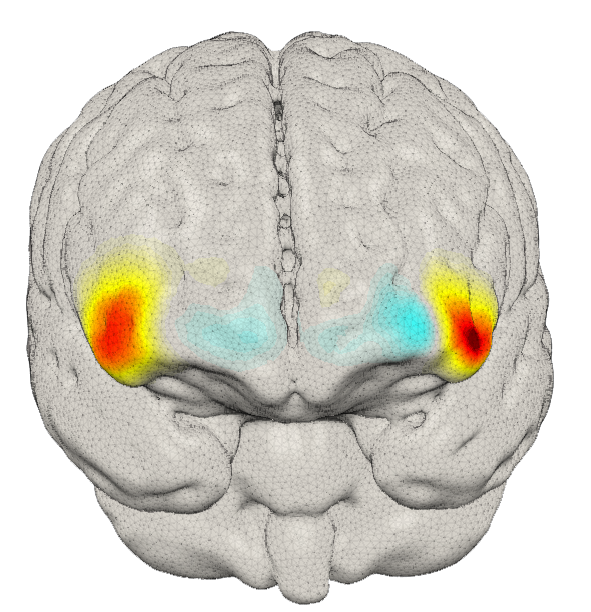
You may have noticed there aren’t any images being generated by fNIRs. This is because in order to generate images you must use Diffuse Optical Tomography (DOT). Simply put, DOT involves using lots of fNIRs sources and detectors and overlapping them to generate an image. Using these images in conjunction with a functional experiment, you can see which areas of the brain ‘light up’ when conducting particular activities. Since fNIRs can be used while moving, the sky is the limit when coming up with experiments to conduct.
The Promising Future of fNIRs/ Reflection
After discovering this promising technology, I felt compelled to write about it. I found its resistance to motion artifacts particularly interesting, as a biomedical engineer, they are something I have to deal with in all of my projects. Additionally, this feature means fNIRs has the potential to investigate infant brains as MRI and fMRI require the patient to stay very still, which can be very difficult for infants and children.
A researcher at the University of Southampton; Dr Ernesto Elias Vidal Rosas, is currently working on an fNIRs system that will tackle the issue of the lower spatial resolution, one of the main drawbacks of the technology. He inspired me to investigate this technology after discovering one of his written papers on the topic.
Personally I see this technology rivalling that of fMRI in its researching capabilities in infants not only due to the motion resistance but also the ability to conduct naturalistic experiments, potentially using technology like VR in order to investigate the brain’s activity when interacting with the world outside the lab. Additionally, the sensor could be used in other areas of the body for example, when placed just below the ribcage, “[fNIRs] has shown promise in being a more accurate, and less bias sensor compared to the gold standard”, Dr Ernesto Elias Vidal Rosas.
This is a good blog. It nicely demonstrates a good understanding of organ-on-a-chip technology and clearly explains its purpose and…
This is a good blog, very engaging with a good backgroud to 3D bioprinting. You could improve your blog with…
This is a good, very interesting blog about necrobotics. It explores the idea of necrobiotics which is fairly new approach…
This is a good blog. You introduce the reader to the topic of prosthetics and bionic limbs in a very…
This is a good blog introducing hernia mesh benefits and drawbacks. You create a narrative in this blog, which showcase…