The University of Southampton

Type 1 Diabetes: The fight for a cure

Imagine if your ability to eat relied on whether you could solve a handful of maths questions, lines of algebra blocking you from simply reaching over to the snack drawer and grabbing a bag of your favourite crisps. Sounds like a nightmare, right? But for 464,000 people in the UK alone, this is a version of their reality whilst living with type 1 diabetes, an autoimmune disease that prevents your body from producing insulin [1].

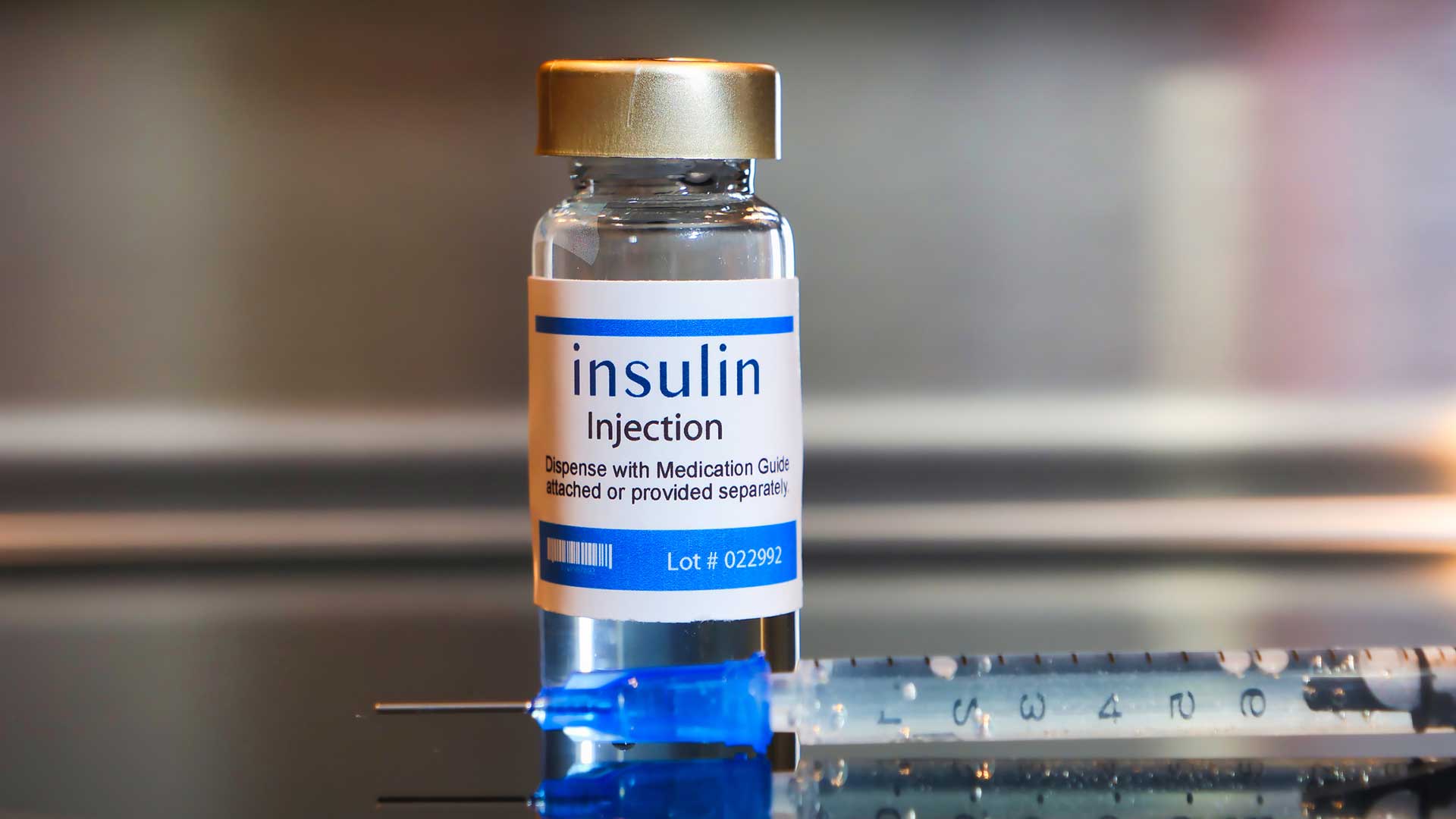
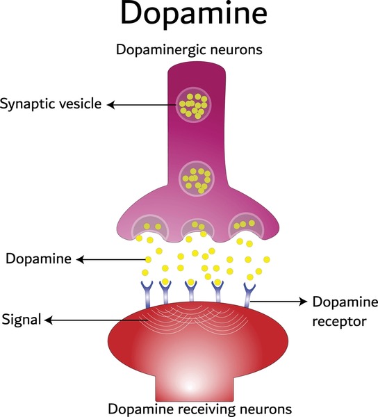
Insulin, a vital hormone produced by the pancreas after the ingestion of food, enables our body to transfer glucose from our blood into our cells, allowing it to be used as energy [2].

Image available at: https://gluroo.com/blog/diabetes-101/insulin-faqs-type-1-diabetes/ (accessed: 09/03/25)

Those with type 1 diabetes have a pancreas that is unable to produce enough insulin to promote this transfer causing their levels of blood glucose to slowly increase with each meal, the sugars predominantly being expelled only as waste via frequent urination. As a result, insulin must be supplemented through injection, quantities being meticulously calculated dependent on what is being eaten and when. Whilst with years of practice calculating the amount of insulin you need can become almost second nature, many with this disease long for a day where they can eat freely, not having to constantly monitor their blood sugar levels and symptoms.

A short video explaining some key aspects of type 1 diabetes – closed captions are available!

Current Technologies

Whilst technology currently exists which aims to make the lives of those with diabetes easier, nothing works quite as well as a pancreas. For example, DexCom is a company which produces a small sensor which allows for the continuous monitoring of blood sugar levels, a connecting app receiving and displaying the statistics on a regular basis. Having an error of only 8.2%, this sensor would appear to be amazing in almost any other application, but when it comes to someone’s health you can never be accurate enough [3].

This video follows a young woman called Jordan and how DexCom allowed her to regain control of her life and love for sport (closed captions available)

With pancreas transplants carrying the same risks as any other major surgery, the possibility of rejection urging healthcare professionals to encourage the majority to manage their condition through diet and medication, most are left with these sensors as their most advanced form of care. Therefore, although some aspects of life with type 1 diabetes can be made more “convenient”, the fight for an accessible cure is still ongoing.

Stem Cells and Ethics

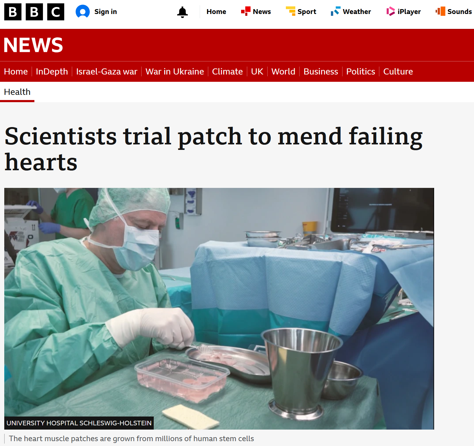
Excitingly, it has been reported that a 25-year-old woman with type 1 diabetes has undergone a stem cell transplant enabling her pancreas to begin producing insulin again, it continuing to do so even a year post transplant – more about this encouraging story can be read here! Whilst this seems promising, the procedure does come with risks. It is believed that a form of immunosuppression is required in order to prevent complications, making the patient more vulnerable to other illnesses and infections.

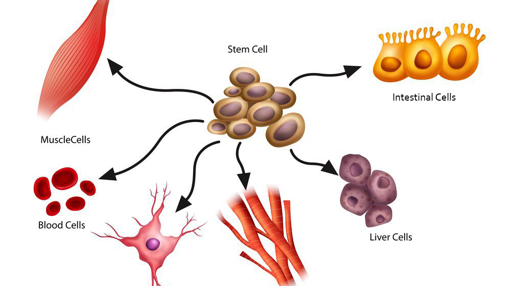
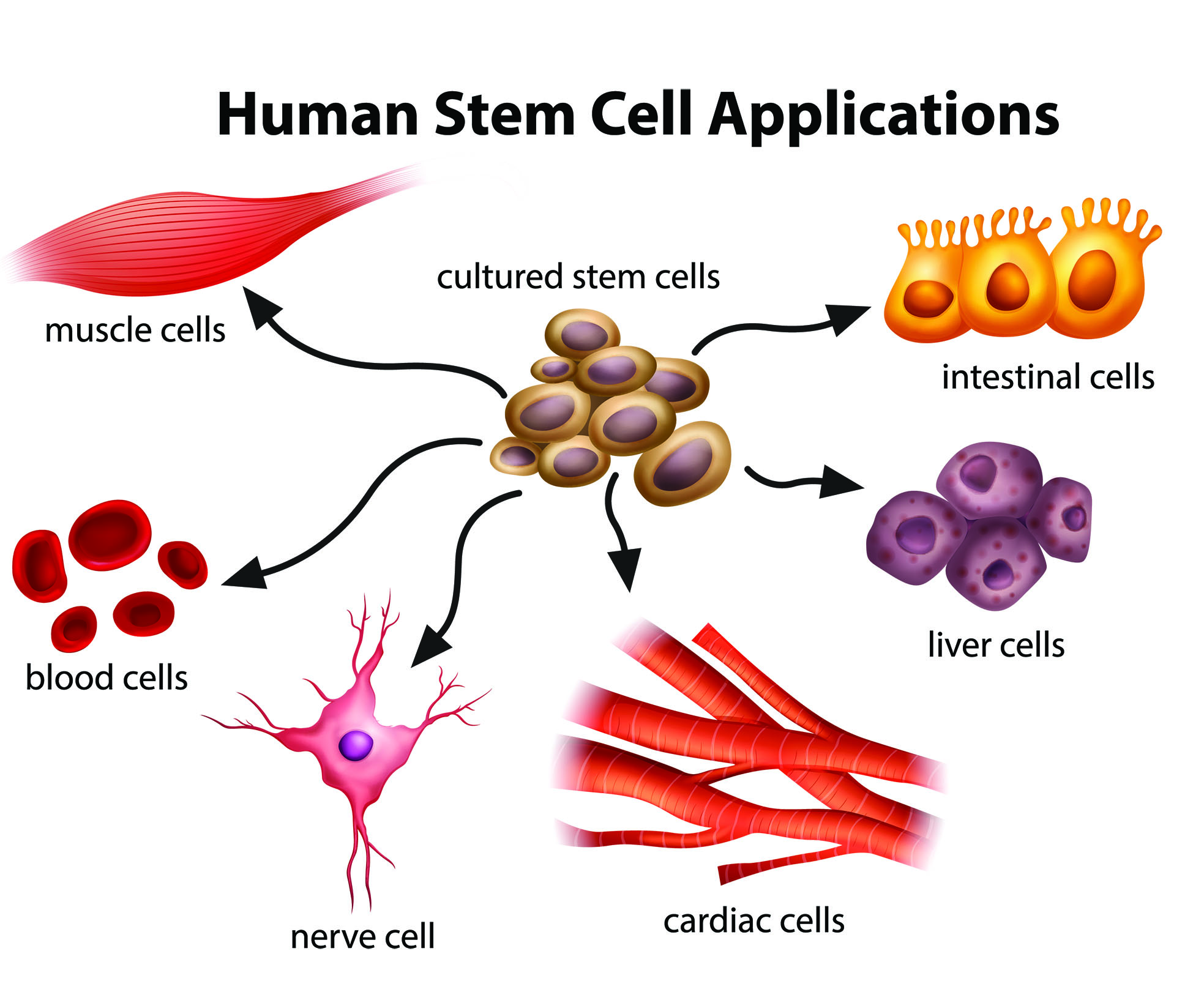
Furthermore, there is currently a large volume of debate over the ethics of using stem cells in treatments. Embryonic stem cells, those that are typically used in such treatments, can only be obtained through the destruction of a human embryo, a process that breeds concern surrounding the sanctity and rights of early human life [4].

Image available at: https://bioinformant.com/what-are-stem-cells/ (accessed: 09/03/25)

Whilst many believe using a potential human life in this manner is unethical, I can’t help but wonder if denying somebody a better quality of life that could be so easily provided is just as cruel. How would you feel if a cure to something that controlled your daily life was just out of reach?

References

[1]Diabetes UK, “How Many People in the UK Have diabetes?,” Diabetes UK, 2024. https://www.diabetes.org.uk/about-us/about-the-charity/our-strategy/statistics

[2] NHS , “What is type 1 diabetes?,” nhs.uk, Nov. 31, 2024. https://www.nhs.uk/conditions/type-1-diabetes/what-is-type-1-diabetes/

[3]S. K. Garg et al., “Accuracy and Safety of Dexcom G7 Continuous Glucose Monitoring in Adults with Diabetes,” Diabetes Technology & Therapeutics, vol. 24, no. 6, pp. 373–380, Jun. 2022, doi: https://doi.org/10.1089/dia.2022.0011.

[4] L. A. Cona, “Stem Cell Research Controversy: A Deep Dive (2023),” www.dvcstem.com, Sep. 14, 2023. https://www.dvcstem.com/post/stem-cell-research-controversy

‌

Cloning: Simply Science Fiction?

The idea that an individual could be identically replicated has captivated popular culture for decades. A clone is defined in biology as an organism or cell, produced asexually from one ancestor, to which they are genetically identical. Appearing in the likes of the Star Wars franchise, Oblivion, and more recently Mickey-17 released just this month, clones in science fiction films are often portrayed in a way that challenges this definition. When reproductive cloning and Dolly the sheep were introduced in Nick Evans’ lecture on stem cells, I asked myself the following questions:

  • To what extent does cloning exist?
  • Why haven’t we cloned humans?
  • What are the applications of cloning?

Animal cloning – Dolly the sheep

On the 5th of July, 1996, the first mammal cloned from an adult somatic cell was born – Dolly the Finn-Dorset sheep. The scientific advancement captured the attention of the media at the time, and continues to do so today (see links on right). The procedure involved somatic cell nuclear transfer, which is a type of reproductive cloning. The diagram below summarises the method.

Image from EncyclopĂŚdia Britannica, Inc.

Dolly essentially had three mothers, with the DNA from the egg cell of a Scottish Blackface sheep being replaced by the DNA from a Finn-Dorset sheep. The resulting hybrid cells were placed in the uterus of another Scottish Blackface, and astonishingly, Dolly was born with identical genes to the nucleus donor sheep. In theory, the exact same procedure could be completed with humans. It should be noted that it took 227 attempts to produce one sheep clone. Even today, mammalian cloning is highly inefficient.


Ethical Considerations

Following the birth of Dolly, the United Nations Educational, Scientific, and Cultural Organisation (UNESCO) immediately banned cloning. It goes without saying that cloning humans would raise innumerable ethical issues. Below are the most notable (click on the arrows for more information):

Eugenics

An early ethical consideration, following John Lederberg advocating for cloning in 1966. He saw it as an appropriate means to improve the human race – cloning certain individuals for their desirable traits. Many see this is a ‘new form of eugenics’, which is considered immoral by most, due to questions of equality and coercion.

Danger

Utilitarianism focuses on the direct outcome, raising the concern that cloning human beings by nuclear transfer is too dangerous. Too many embryos in animal cloning experiments expire or result in miscarriage.

Individuality

Many believe that cloning violates our dignity, uses people as means, affronts our uniqueness, and threatens our humanity.

Embryonic research

To even research human cloning, a huge amount of human donated eggs would have to be acquired by scientists. The nature of the nuclear transfer method, with implantation would also require the research to surpass the 14-day limit of embryo experimentation, raising questions on identity and the value of human life.


Therapeutic Cloning

Therapeutic cloning is a practical method which in my opinion avoids the ethical issues raised above. It also involves nuclear transfer, but the cells are never implanted. Instead, the embryo is cloned to produce stem cells. Since these would be genetically identical to the donor, these stem cells are less likely to be rejected by the patient.

Image from Mitalipov Lab/OHSU

Conclusion

It seems to me that human cloning is inefficient, immoral, and mostly pointless. The replicas we see in science fiction don’t reflect biology, and with therapeutic cloning, there is no need to clone entire humans for treatment.

“Your scientists were so preoccupied with whether or not they could, they didn’t stop to think if they should.”

Ian Malcolm, Jurassic Park (1993)

3D Bioprinting

Press a button, print an organ. Perhaps the idea is not as Sci-Fi as it sounds. 3D bioprinting is an advancing field that uses layer-by-layer positioning of cells and biomaterials to produce living scaffolds for a variety of uses.

Depending on the qualities needed in the final product, techniques and materials vary. All start with a digital 3D model produced using computer aided design (CAD) software. One powerful feature of this is that CT and MRI scan data from patient can be used to tailor, for an example a tracheal graft, to personalised dimensions.

Next, the structure can be bioprinted using a bioink. Things to consider when choosing a bioink are the cells, biomaterial and growth factors included. Biomaterials such as alginate and polyvinyl-alcohol provide a structural scaffold for new cells to grow on. The ideal biomaterial must be biocompatible (not trigger an immune response) and biodegrade at the same rate as the new tissue grows.                                     

Cells and growth factors goes hand in hand, to print a bone graft you would use mesenchymal stem cells and a corresponding growth factor or chemical that encourages the pathway of differentiation looked for. The bioink used can dictate the printing method. Three popular approaches include inkjet-based, laser assisted and extrusion-based bioprinting. Each has their own set of opportunities and challenges.

Inkjet bioprinting uses the bioink droplets deposited by a piezoelectric actuator. Laser-assisted uses an a donor layer than absorbs energy from laser stimulation with a bioink layer underneath it and a collecting layer to form the tissue constructs. Extrusion uses mechanical force to deposit a continuous cylindrical stream of bioink.
Mechanisms of 3D bioprinting: Adapted from an image by Loai et al, licenced under Creative Commons Attribution 4.0 International License from https://doi.org/10.20900/rmf20190004

Droplets of bioink, generated either through heating or a piezoelectric effect, are deposited on the substrate. High throughput, high resolution, low expense and strong cell viability count in favour of this technique. The key drawback is the need for low viscosity bioink to pass through the fine nozzle which results in low structural quality.

If a strong structure is a priority then extrusion printing comes to the fore. It relies on pneumatic or mechanical force to print uninterrupted lines of a far more viscous bioink. However, the increased shear stress involved reduces cell viability. High viscosity bioinks tends to result in lower resolution and the process is much slower.

This uses a laser induced forward transfer technique (LIFT) which means that a laser is used to force material from a thin donor layer of bioink onto the substrate being printed onto. Advantages of this include high cell viability as there is no direct contact between the bioink and the dispenser so no stress forces and that the technique is compatible with a wide range of bioink of varying viscosities. Unfortunately, the complexity of this method results in high equipment cost.

Potential Uses

  • Organ replacement
  • Tissue grafts such as skin, bone muscle and cartilage
  • Organs on a chip – used for drug and vaccination screening
  • Drug delivery scaffolds
  • Building disease models

Printing organs for those currently awaiting a donor remains science fiction for now but great progress is being made towards that goal. However, even this is just the start as ethical considerations need to be made.

Integral to the whole process are stem cells and their source. The ethics of stem cells use are hotly debated but the alternative, induced pluripotent stem cells, are associated with more unpredictable behaviour.

Safety and quality control of bioprinted organs need to be considered, and this is made harder personalisation of treatment which reduces replicability of results. Arguably, for a terminally ill patient the benefits of an untested treatment outweigh the risk, but can this then be applied to non-life-threatening cases?

A New You! Stem Cells in Regenerative Medicine

Can the body heal itself? The power of stem cells in a new era of healing

Stem Cells are cells that can self-renew and differentiate, this is crucial for the development of an organism as well as repair after injury (Mayo Clinic, no date). As a cornerstone of regenerative medicine, they offer innovative new solutions to treating diseases with limited treatment options such as restoring damaged organs and tissues (Wang et al. 2024).

Understanding Stem Cells

It is important to understand the different types of stem cell

  • Multipotent: The ability to differentiate into more than one cell type in the body
  • Pluripotent: The ability to differentiate into all of the various cell types in the body

Pluripotent and multipotent stem cells originate from different sources. Pluripotent stem cells are derived from early-stage human embryos, these are capable of dividing without differentiation and can develop into the primary three germ layers. In comparison, adult stem cells can only differentiate into the cell type of the tissue that they are found (Mayo Clinic, 2025). Researchers have developed an induced pluripotent stem cell, similar to embryonic stem cells, but, formed by transferring embryonic genes to a somatic cell (Mayo Clinic, 2025).

What is Regenerative Medicine

Regenerative medicine is a branch of medicine that focuses on healing or replacing organs and tissues damaged by factors such as disease or trauma. ‘Healing’ is achieved by replacing missing tissue structurally or functionally using stem cells such as mesenchymal stem cells induced pluripotent. This is achieved through materials as well as de novo-generated cells. It is also possible to leverage the body’s inate healing response, however humans lack regenerative capacity (Mao and Mooney, 2015). Regenerative medicine has the potential to treat neurodegenerative diseases as well as heart failure, for example.

Stem Cells in Regenerative Medicine

Adult stem cells such as mesenchymal and induced pluripotent stem cells (iPSCs) , exhibit multilineage differentiation capacities as well as immunomodulatory properties (Li, Luanpitpong, and Kheolamai, 2022). There have been successful treatments using these stem cells for bone and cartilage regeneration as well as spinal cord injuries and diabetes (Hoang et al., 2022). allowing them to differentiate into required tissues and treat injuries, inflammation, and age-related disorders through regeneration of muscle, cartilage, and muscle regeneration. Human pluripotent stem cells (hPSCs), embryonic stem cells (ESCs), and (iPSCs) can differentiate into any cell type so have a very high potential (Wang et al., 2024).

Challenges Associated

However, there remain risks associated with stem cell use such as a heightened risk of tumor formation, and immune rejection, and it’s not guaranteed that the cells will survive post-translation.

The treatments with the most therapeutic potential use embryonic stem cells, due to their pluripotency. However, the obtainment of ESCs is controversial due to the destruction of human embryos, many people object to religious and ethic principles (Margiana et al., 2022). Stem cell therapies also have a high cost meaning availability is restricted to wealthier patients (Hoang et al., 2022).

Conclusion

The advancement of regenerative medicine presents exciting opportunities such as within genetic modification, combining stem cells with drug delivery as well as biomaterial scaffolds (Li, Luanpitpong, and Kheolamai, 2022). However, currently stem cells remain at the forefront for conditions such as cardiovascular disease and organ regeneration. However, further research is needed to combat concerns such as immune rejection and tumor risks. For this treatment to become mainstream steps will have to be taken to address high costs and accessibility issues as well as the need for a global regulatory body to monitor its use.

Bibliography

Hoang, Duc M., Phuong T. Pham, Trung Q. Bach, Anh T. L. Ngo, Quyen T. Nguyen, Trang T. K. Phan, Giang H. Nguyen, et al. ‘Stem Cell-Based Therapy for Human Diseases’. Signal Transduction and Targeted Therapy 7, no. 1 (6 August 2022): 1–41. https://doi.org/10.1038/s41392-022-01134-4.

Li, Jingting, Sudjit Luanpitpong, and Pakpoom Kheolamai. ‘Editorial: Adult Stem Cells for Regenerative Medicine: From Cell Fate to Clinical Applications’. Frontiers in Cell and Developmental Biology 10 (31 October 2022). https://doi.org/10.3389/fcell.2022.1069665.

Mao, Angelo S., and David J. Mooney. ‘Regenerative Medicine: Current Therapies and Future Directions’. Proceedings of the National Academy of Sciences 112, no. 47 (24 November 2015): 14452–59. https://doi.org/10.1073/pnas.1508520112.

Margiana, Ria, Alexander Markov, Angelina O. Zekiy, Mohammed Ubaid Hamza, Khalid A. Al-Dabbagh, Sura Hasan Al-Zubaidi, Noora M. Hameed, et al. ‘Clinical Application of Mesenchymal Stem Cell in Regenerative Medicine: A Narrative Review’. Stem Cell Research & Therapy 13, no. 1 (28 July 2022): 366. https://doi.org/10.1186/s13287-022-03054-0.

Mayo Clinic. ‘Answers to Your Questions about Stem Cell Research’. Accessed 5 March 2025. https://www.mayoclinic.org/tests-procedures/bone-marrow-transplant/in-depth/stem-cells/art-20048117.

Mayo Clinic. ‘Answers to Your Questions about Stem Cell Research’. Accessed 5 March 2025. https://www.mayoclinic.org/tests-procedures/bone-marrow-transplant/in-depth/stem-cells/art-20048117.

Wang, Jipeng, Gang Deng, Shuyi Wang, Shuang Li, Peng Song, Kun Lin, Xiaoxiang Xu, and Zuhong He. ‘Enhancing Regenerative Medicine: The Crucial Role of Stem Cell Therapy’. Frontiers in Neuroscience 18 (8 February 2024). https://doi.org/10.3389/fnins.2024.1269577.

Petri Dish to Patient : How 3D Bioprinting is Changing Transplants

“Why can’t we just print more organs?” Overcoming the organ shortage crisis with Bioengineering

Globally, the demand for organ transplants exceeds supply. In the United States as of 2021 116,566 patients were waiting for an organ transplant, 6 564 patients died waiting for an organ. Despite this, only 41 354 transplants were performed (Kupiec-Weglinski 2022). 3D bioprinting is an emerging technology that offers a promising solution by creating bioengineered organs and tissues, reducing patients left waiting for life-saving treatments and reliance on human donors (Bose, 2023)

3D bioprinting is an innovative technology that involves printing layer by layer, combining biomaterials, bio-inks, and living cells (Panja et al., 2022). This provides an opportunity to engineer human tissues and organs for medical use such as :

  • Drug testing on human models (more reliable and reduces demand for animal models)
  • Custom implants
  • Reducing the waitlist for replacement human organs

How It Works

There are 4 main bioprinting techniques

Technique How it Works Pros Cons
Extrusion Bioprinting Uses mechanical force to push bio-ink through a nozzle Good for large tissues
Can use high-viscosity bio-inks
Can cause damage to cells from pressure
Laser-Assisted Bioprinting Uses lasers to deposit biomaterials High precision
No nozzles needed
Expensive, Slow
Stereolithography (SLA)Uses UV light to harden layers of biomaterials High resolution, Fast Limited material options
Inkjet Printing Droplet-basedFast, Cost effective Low precision
Can only use low-viscosity bio-inks

(Panja et al., 2022)

Applications

Recently there have been significant breakthroughs in replicating complex human organs especially hollow organs such as the lungs, heart, and digestive system. Despite the immense progress, technical challenges still persist.

Bioprinting Lungs 

  • Lung diseases such as COPD and COVID-19 have increased demand for lung transplants. The focus remains on the airways and alveoli.  Advancements have been made in the development of artificial alveolar models using inkjet printing, hydrogels, and synthetic polymers.  Despite this, these have not been applied to organ replacement yet (Panja et al., 2022). Achieving the highly complex structure and function of alveoli is still a significant hurdle. 

3D Printed Heart Tissue 

  •  A heart has been bioprinted including blood vessels. This is crucial when developing the functionality of replacement organs. This was achieved using a technology called Coaxial Sacridicial Writing in Functional Tissue. This adds layers of real blood vessels, blood supply is essential for sustaining all bioprinted organs (Brownell, L. 2024).

Digestive System 

  • Researchers have also seen developments in the bioprinting of the digestive system, including stomach, intestines and bile ducts. There is enormous potential for this to revolutionize drug testing and reduce reliance on animal models. However, barriers remain due to the inability to replicate the mechanical processes of the digestive system such as peristalsis(Panja et al., 2022. 


Challenges and Ethical Concerns

  • Cost: Bioprinting is an expensive technology, with significant costs from research, development, and clinical trials. As well as the extensive laboratory equipment, this means that the early technology is likely to be limited to the wealthy (Bose, P., 2023).
  • Tissue Vascularisation: functional blood vessels are imperative for delivering oxygen and nutrients, and keeping tissues alive. Functional blood vessels are challenging due to their microscopic size and complexity (Panja et al., 2022).
  • Regulation and Safety: international regulations are necessary to ensure safety, efficacy and ethics have been taken into consideration (Brownell, L. 2024).

Conclusion

3D printing has the potential to make fully functional, transplantable organs a reality. There needs to be a focus on personalization to reduce the risk of elimination. However, vascularisation will remain a significant barrier to applicability. However, the potential to remove donor waitlists and animal models.

Could we be entering an era of on-demand organ transplants?

Bibliography

Bose, P. (2023). Bioprinting Organs: A Look into the Future of Transplantation. News-Medical. Available at: https://www.news-medical.net/health/Bioprinting-Organs-A-Look-into-the-Future-of-Transplantation.aspx [04/03/25].

Brownell, L. (2024). 3D-printed blood vessels bring artificial organs closer to reality. Harvard John A. Paulson School of Engineering and Applied Sciences. Available at: https://seas.harvard.edu/news/2024/08/3d-printed-blood-vessels-bring-artificial-organs-closer-reality [04/03/25].

Panja, N., Maji, S., Choudhuri, S., Ali, K. A., & Hossain, C. M. (2022). 3D Bioprinting of Human Hollow Organs. AAPS PharmSciTech, 23(5), p.139. Available at: https://doi.org/10.1208/s12249-022-02279-9 [04/03/25].

Kupiec-Weglinski, Jerzy W. ‘Grand Challenges in Organ Transplantation’. Frontiers in Transplantation 1 (6 May 2022). https://doi.org/10.3389/frtra.2022.897679. [04/03/25]

Knee replacements: Cost for Mobility

“We don’t appreciate what we have until it’s gone.” This phrase resonated deeply with me during the lecture on joint replacements. We often take for granted the ability to move and stand without pain—until one day, we can’t. Millions of lives have been transformed by the invention of knee replacement, restoring people’s movement but also their independence. But what are the challenges of these replacements and what does their future look like?

Evolution of knee replacements

The idea of replacing damaged joints isn’t new and has been around for hundreds of years where ancient civilisations experimented with rudimentary prosthetics. Modern knee replacement surgery, or total knee arthroplasty (TKA) however, only emerged in the mid-20th century. Advances in materials, surgical precision, and rehabilitation have meant that patients today can regain almost all lost function. Whilst these improvements have been greatly beneficial, there are still risks and limitations involved.

image: Fixed vs mobile bearing of knee replacements https://link.springer.com/chapter/10.1007/978-981-16-8591-0_3

More Than Just a Mechanical Problem?

Engineering is at the core of a knee replacement where surgeons remove damaged bone and replace it with metal and synthetic components that are specifically designed to mimic the joint’s natural function. The real challenge however, is in how the body allows the artificial joint to work within our bodies, where ligaments, tendons, and muscles must adjust to the artificial joint, and imbalances in force distribution can lead to complications such as implant loosening. The risk of rejection is also prevalent as the body may recognise the implant as foreign, triggering low-grade inflammation that can affect the implant’s longevity.

As with any surgery there is the emotional and psychological side. How does it feel having to relearn simple movements such as walking? Regaining autonomy can be life-changing, but rehabilitation can be long and demanding.

Challenges and Ethical Considerations

Despite its success and widespread use, it still has its limitations.

  • Longevity of implants: A knee prosthesis only lasts 15–20 years and so younger patients may require multiple complex surgeries.
  • Access to prosthetics: Not everyone can afford a knee replacement. Is it right that orthopaedic treatments are more readily available to those with financial means?
  • Surgical risks: Infections and rejection are still major risks.

In a recent BBC article from last year, Heather considered herself “lucky” to have had her left knee replaced after two years of pain. This article highlights a wider systemic issue in healthcare-availability. The question remains as how can we solve this?

This shows that there is still a long way to go when talking about this type of technology and highlights the need for continuous innovation in this area.

Future of Knee Replacements: Can We Do Any Better?

With recent advancements in areas such as 3D printing and robotic-assisted surgery, the future of knee replacements is still promising. One day, could we use cartilage grown in a lab to produce personalised implants? And could AI-driven rehabilitation programs increase patients’ recovery outcomes?

This lecture changed the way I think about joint replacements as a field that intersects engineering, biology, and ethics. It made me think about the the privilege of mobility and the importance of continuous innovation in medical technology.

A Reflection on Human Head Transplants

Following discussion on organ transplants, my reading led me to the debate of head transplantation, which, to those with terminal disease but a healthy head and brain [1], has the potential to extend life. It would be a last-resort treatment for patients whose body is affected but whose mind and head are healthy, hence treating conditions such as quadriplegia, progressive diseases, and inoperable cancers that have not extended to the brain [2]. In 2019, Valery Spiridonov—a man with Werdnig-Hoffmann disease, a muscle-wasting condition—pulled out of what would have been the first attempted human head transplant [3].

The surgery would require an immunologically matched brain-dead donor with a healthy body, onto which the head of the patient can be transplanted. The procedure has been performed on animals without long-term success [4], and in 2017, it was completed on a human cadaver [5]. Gkasdaris et al. discussed human head transplantation and highlighted the surgical issues involved as donor body selection, head and body interventions on the recipient and donor, ischemia time (managing the blood flow between the head and donor body), spinal fusion and spinal cord reattachment, and post-operative issues [1]. Beyond the surgical hurdles, ethical concerns are raised; notably, the EANS ethico-legal committee concluded that it is ethically unacceptable to attempt a head transplant [6]. Literature considers the great risk involved during and after surgery and the reality of living with the body of another person; for example, a 2017 study by Wolpe considers the extent to which our body, alongside the brain, “makes us who we are” [3]. Legality must also be taken into account. Wolpe points out that the surgery involves intentional decapitation, which means that, by definition, the death of a patient would be murder.

The idea of a head transplant is one that we, as the general public, expect to see in science fiction media, and would therefore view as something detached from reality. Frankenstein’s monster is often referenced in this discussion. We would consider the consequences of such a narrative—written to shock and scare us—and conclude that this is not something that should happen in ‘real life’. We have been told by the fiction we grow up around to be wary of the scientific unknown and the ‘mad scientist’ archetype who would attempt something so shocking and seemingly without consideration of ethical questions. It is difficult to suspend these preconceived ideas of what should and should not be done, and dull the instinctive, fearful dismissal of an attempt. Just the phrase ‘head transplant’ elicits shock, even as somebody so used to hearing about extreme surgical procedures. Through reading and consideration, I hope to form my own opinion on the subject, beyond general concern and morbid fascination.

I believe that the surgical challenges are the lesser issue when it comes to the feasibility of head transplants. Surgical technique is rapidly advancing and there will come a point in medical knowledge where every challenge associated with the procedure will be surmountable. My reading found this to be an existing approach in the conversation; Spagnolo et al. says, in a paper about the possibility of head transplants, “Despite the uncertainty regarding the technical feasibility of this procedure, for the sake of argument, we will assume that the procedure is possible and feasible to perform” [7]. Even so, the first attempt will come with great risk; if we reached a point where an attempt could be made, should it?

Whilst no comparison can be made to head transplants, this scenario brings to mind the case of Ladan and Laleh Bijani: conjoined twins who both died during a separation attempt on 8 July 2003, at twenty-nine years old. The surgery, which separated their heads, had been denied in 1996 based on high risk [8], but was accepted by another surgical team in 2003 despite major safety concerns. Varying reports exist on which parties involved agreed that the surgery should go ahead, but both twins made clear their desire for the surgery by explaining that their lives conjoined “were worse than death”. Similarities can be drawn between this case and the scenario of a person who would undergo head transplantation to avoid a fate they regarded to be ‘worse than death’. To form an opinion on the head transplant debate, I considered the question of whether Ladan and Laleh’s separation surgery should have been performed, knowing the outcome of it. Can the blame for their deaths be placed on the surgeons, or the twins? Perhaps societal pressure—and the public spectacle of such a novel surgery, with the relations between Singapore and Iran affected as a consequence [9]—could have contributed to the decision to proceed with the surgery? Hence, was it correct to place such a spotlight on the case, and would the first head transplant be subject to similar public interest that could influence the decisions of the parties involved?

It is my opinion that the correct choice was made in respecting the wishes of the twins. We will once day live in a world where the knowledge will exist to attempt the first human head transplant. There must always be a first, and that first will be performed on a patient who, like Ladan and Laleh, see their current condition as one worth risking death to escape from. Once it can be attempted, then, even with all the concern that remains, I believe that it should. Still, the only conclusion I can confidently draw from this can be summed up by Gkasdris et al.: “The scientific community should not consider [human head transplantation] as a product of imagination anymore” [1].

References:

[1] G. Gkasdaris and T. Birbilis, “First Human Head Transplantation: Surgically Challenging, Ethically Controversial and Historically Tempting – an Experimental Endeavor or a Scientific Landmark?,” Mædica, vol. 14, no. 1, pp. 5–11, Mar. 2019, doi: https://doi.org/10.26574/maedica.2019.14.1.5.

[2] B. Peters, “What to Expect From a Head Transplant,” Verywell Health, Jun. 08, 2022. https://www.verywellhealth.com/head-transplant-4801452

[3] Spinalcord. com Team, “Warning Signs of a Serious Spinal Contusion,” Spinalcord.com, Dec. 03, 2020. https://www.spinalcord.com/blog/russian-man-volunteers-to-be-the-first-full-head-transplant (accessed Mar. 06, 2025).

[4] P. R. Wolpe, “Ahead of Our Time: Why Head Transplantation Is Ethically Unsupportable,” AJOB Neuroscience, vol. 8, no. 4, pp. 206–210, Oct. 2017, doi: https://doi.org/10.1080/21507740.2017.1392386.

[5] X. Ren et al., “First cephalosomatic anastomosis in a human model,” Surgical Neurology International, vol. 8, no. 1, p. 276, 2017, doi: https://doi.org/10.4103/sni.sni_415_17.

[6] J. Brennum, “The EANS Ethico-legal Committee finds the proposed head transplant project unethical,” Acta Neurochirurgica, vol. 158, no. 12, pp. 2251–2252, Oct. 2016, doi: https://doi.org/10.1007/s00701-016-2986-y.

[7] A. Cartolovni and A. Spagnolo, “Ethical considerations regarding head transplantation,” Surgical Neurology International, vol. 6, no. 1, p. 103, 2015, doi: https://doi.org/10.4103/2152-7806.158785.

[8] N. Ahmad and L. Board, “World’s first separation of adult Siamese twins in Singapore,” Nlb.gov.sg, 2023. https://www.nlb.gov.sg/main/article-detail?cmsuuid=8909f20d-50e5-43f3-86ee-0fe93f58e0a1 (accessed Oct. 08, 2024).

[9] Wikipedia Contributors, “Ladan and Laleh Bijani,” Wikipedia, Jan. 12, 2025.

‌

‌

‌

A Heart Alternative – How Far Can We Go?

Blog audio transcript:

Following the anatomy lab visit and lectures on stem cells, tissue engineering and prosthetics, it made me think about how far we can go towards developing an organ from a non-human origin. The heart stood out as one that could be in the realms of possibility compared to some of the more complex organs.

A labelled Diagram of the Internal Anatomy of the Heart
A Labelled Diagram of the Internal Anatomy of the Heart. Available at https://commons.wikimedia.org/wiki/File:2008_Internal_Anatomy_of_the_HeartN.jpg (Accessed: 25/02/2025). By OpenStax College, licensed under the Creative Commons Attribution 3.0 Unported license from Wikimedia Commons.

Genetically modified pig heart transplants have been carried out, with the first transplant performed in 2022 on a 57-year-old individual with end stage heart failure. This operation, formally called xenotransplantation, worked well for seven weeks, however the patient passed with heart failure after this time. It was found that the heart was vulnerable to rejection, it also contained traces of a virus that infects pigs.

A BBC Video Reporting on the First Patient to Receive a Pig Heart Transplant (Accessed: 23/02/2025). Captions are available on this video.

There is a very good reflective article linked here which talks about where to go after this xenotransplantation was carried out. Feel free to have a look if you are interested in reading more.

This transplant brings up ethical points to consider along with scientific challenges:

  • To what extent is it right to sacrifice animals for the benefit of humankind? Applying ethical concepts, most people would say more good is done by saving a human life than by sacrificing an animal such as a pig. Furthermore, if a pig could be genetically modified to become more humanised in the future, then perhaps more than one human life could be saved for each pig. Provided that the pig is well cared for during its lifetime, then this action of using its tissue would overall be deemed as “good” and the “right” action.
  • Scientific problems can happen during any transplantation. Specifically for pig heart xenotransplantation, immediate and acute rejection of the tissue can be an issue. Thrombosis, blood clots, can also occur due to a haematological incompatibility.

This video summarises the scientific and ethical implications of xenotransplantation:

An Interview Discussing the Scientific and Ethical Challenges Around Xenotransplantation (Accessed: 23/02/2025). Captions are available on this video.

A recent BBC article published in January 2025 highlighted work done in Germany where stem cells were used to develop patches of heart muscle cells which were then grafted onto damaged tissue. This could help patients with heart failure, giving encouraging results in trials.

A Screenshot of a BBC Article Titled “Scientists trial patch to mend failing hearts”
A Screenshot of a BBC Article Titled “Scientists trial patch to mend failing hearts”. Available to read at https://www.bbc.co.uk/news/articles/cgkjnl0dve8o (Accessed: 24/02/2025).

From a societal perspective, most of these developments on repairing heart tissue are novel. A high element of risk is not always worth it; however, it might be for those who have little or no alternative.

It is not uncommon to regularly come across news stories regarding the organ shortage. The UK organ donation law changed in May 2020 to an opt-out system meaning that by default adults are considered as potential donors.

In my view the moral status of a human life outweighs the sacrifice of an animal such as a pig. Scientific advance in this area should be encouraged within the framework of ethical boundaries. I cannot find a meaningful downside to pursue alternative methods of sourcing valuable organs.

Stem Cells and Parkinson’s: A New Hope for Treatment?

Parkinson’s disease is a neurogenerative disorder caused by the loss of nerve cells in a part of the brain called substantia nigra. Losing these cells causes a drop in the abundance of a vital chemical called dopamine, leading to a whole host of symptoms, including tremors, stiffness, and dementia. Parkinson’s disease can also strongly impact the mental health of affected people, often associated with depression and anxiety.

This disease is common, affecting more than 1 in 50 people over the age of 65, with some people even experiencing symptoms under 40 years old. The cause of the degeneration of specific nerve cells associated with Parkinson’s disease is unknown, but there is evidence for some new approaches for its treatment using stem cells.

How could stem cells be used?

First, what are stem cells? There are a few kinds of stem cell, but the term generally refers to cells that are capable of developing into other types of cell. Embryonic stem cells, in particular, are pluripotent, meaning they can give rise to all cell types. Induced pluripotent stem cells (iPSCs), which also differentiate into any cell type, can be created via the reprogramming of existing adult cells. Of all stem cells available, these two seem to be the most promising.

One major therapy which has the potential to treat Parkinson’s disease is “Bemdaneprocel”, or “BRT-DA01”. In this therapy, human embryonic stem cells are converted into dopamine-producing neuron progenitors in the lab before being surgically implanted into a specific area of the brain. These cells may be recognised as foreign, so patients must take immunosuppressants for a year. This method began a Phase 1 trial in 2021. “BRT-DA01” showed high safety and potential clinical benefits, and is expected to undergo a Phase 3 trial in the first half of this year.

Another experimental therapy for Parkinson’s disease which aims to replace lost dopaminergic neurons was approved for a Phase 1/2a trial in the US by the FDA in late 2023. In this method (“ANPD001”), skin cells are collected from a patient and reprogrammed in the lab, producing iPSCs which are then converted into immature dopamine neuronal precursor cells. Finally, they are transplanted into the brain, in the hope that they will develop into those crucial neurons. One key advantage of “ANPD001” over “BRT-DA01” is that iPSCs are derived from the patient, so immunosuppressants aren’t needed. As of January this year, this method has been safely carried out with two patient cohorts.

Some ethical issues

While embryonic stem cells may be an effective source for the treatment of Parkinson’s, testing and using them for science raises some interesting ethical questions. These cells are derived from early-stage embryos, in a process involving the destruction of the embryo. The moral status of the embryo in comparison to human life therefore must be questioned. Furthermore, embryos used for scientific purposes are often donated by people undergoing in vitro fertilisation. I feel it is crucial that donors are entirely aware of how their embryos will be used, and clear consent is given.

This is obviously a difficult ethical dilemma, though there seems to be a work-around. Fortunately, iPSCs avoid the ethical questions faced by embryonic stem cells, because they are not derived from embryos. As a result, these cells seem to be a far more acceptable choice for the treatment of Parkinson’s disease, especially from the standpoint of public concern.

Hello!

Hi everyone, I’m Jess and I’m a second year biomedical sciences student. I’m excited to learn more about bioethics and also to be introduced to bioengineering 🙂 I couldn’t attend the workshop today so had to use the panopto recording that had a video about vocal techniques playing over the audio…

I hope I’m doing this right! I guess I learned something about singing today at least 😂