Previous to our lectures on bioethics, I believed that I had a very uncompromising way of assessing scientific ethical concerns: with some exceptions, the advancement of science was worth the cost of a few. From someone who grew up surrounded by scientific minds, this utilitarianism made sense. Why shouldn’t we strive for “the greatest happiness of the greatest number” as stated by Jeremy Bentham? However, since learning about watershed moments in history like the Nuremberg trials, I have learnt that not everything can be so easily segregated into right and wrong. This lead me to reflect on a scientific controversy that I had learned of in my previous year of study: HeLa cells.
What are HeLa cells?
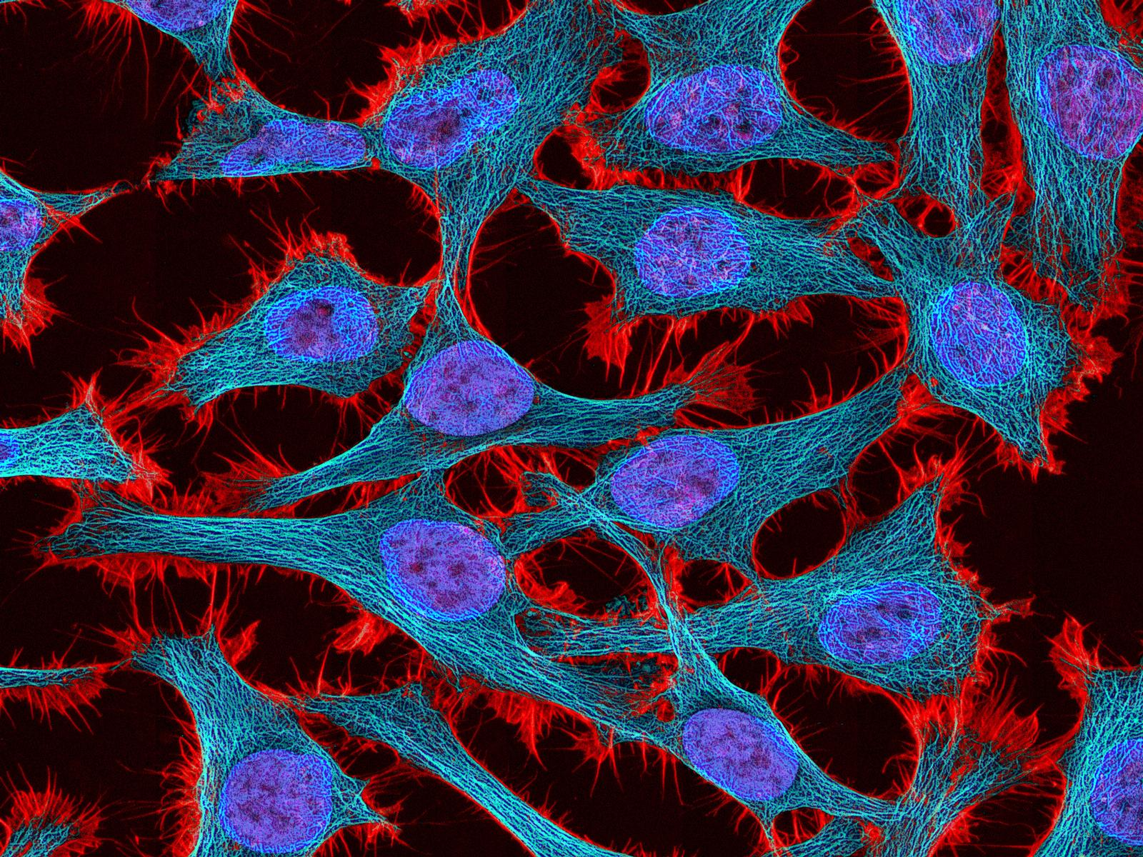
HeLa cells were the first immortal human cell line to be created. Derived from cervical cancer cells, they were cloned in 1953 and freely given to researchers for use in labs. Since then, they have been at the forefront of many medical breakthroughs: from gene mapping to cancer research. Arguably, their most notable use was in the development of the Polio vaccine in 1952.
The Woman Behind The Cells
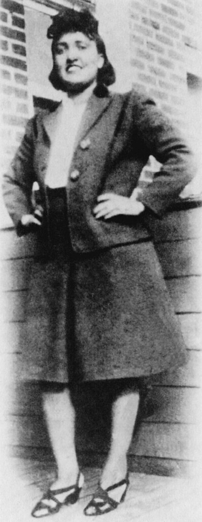
Henrietta Lacks was an African-American woman born in August 1920. She died of an aggressive cervical cancer at the John Hopkins hospital at the age of 31. Unbeknownst to her, one of the gynaecologist who performed her biopsies, Dr Grey, had removed a sample of the tumour for research purposes. He was able to isolate the cancer cells and thus create the first immortal human cell line: HeLa. Knowledge about the use of her cells was not revealed to the family until an article published in Rolling Stone until 1976. Since then, there has been much conversation about the unethical obtainment and distribution of Henrietta’s cells.
Learning from the Past
The main issue regarding HeLa cells was lack of consent. Although this was acceptable at the time, since then, Henrietta’s name has been shared alongside her medical record and even her cell’s genome. Following this, the Henrietta Lacks Foundation, established in 2010, provides grants to descendants, and families whose bodies have been used for research without consent. Henrietta’s descendants have worked tirelessly to create rules over the use of HeLa cells but there is still work to be done. Suggestions of revisions to the NIH Common Rule to protect human participants in US government funded research regarding consent have been put forward. In addition, there is a general consensus that institutions that have used HeLa cells must examine how they will right histories previous wrongs.
What are my Thoughts?
From a scientist’s perspective, the actions of Dr Grey seem understandable. To some, even justifiable. To me however, it is a clear example of where our thirst for knowledge clouds our judgment on what is morally right. Informed consent is a relatively easy standard to meet when obtaining samples for research, especially in cases such as these where the patient is of sound mind. I do not condone the actions of Dr Grey. Despite this, the argument could be made that to remove or limit both past and future contributions of the HeLa cells to modern medicine would be both a disservice to Henrietta. Without her, many scientific advancements would not have been possible. Instead, I think it possible to find a middle ground. One which allows the continued use of the HeLa cells in research whilst also acknowledging their origins.
So reader, over to you. What are your thoughts on the continued use of these cells?
Learn More
If you’re interested in Henrietta Lack’s story, you can find out more with these links below:
- An article on BBC Bitesize on how the HeLa cells have shaped science
- Rebecca Skloot’s book: The Immortal Life of Henrietta Lacks
- The HBO film: The Immortal Life of Henrietta Lacks

This is a good blog. It nicely demonstrates a good understanding of organ-on-a-chip technology and clearly explains its purpose and…
This is a good blog, very engaging with a good backgroud to 3D bioprinting. You could improve your blog with…
This is a good, very interesting blog about necrobotics. It explores the idea of necrobiotics which is fairly new approach…
This is a good blog. You introduce the reader to the topic of prosthetics and bionic limbs in a very…
This is a good blog introducing hernia mesh benefits and drawbacks. You create a narrative in this blog, which showcase…