The University of Southampton

The Science of Eternal Youth: Are Stem Cells the Key to Anti-Aging?

Aging is an inevitable part of life, but what if we could slow it down – or even reverse it? Scientists are exploring the potential of stem cells to unlock the secrets of aging, offering the exciting possibility of longer and healthier lives. Through lectures and discussions, I’ve come to appreciate how stem cell research pushes the boundaries of longevity. But how realistic is this, and what role do stem cells play in the pursuit of eternal youth?

Reactive oxygen species (ROS) are generated by environmental and internal factors like radiation, pollution, and metabolism. ROS cause damage to mitochondrial and nuclear DNA, leading to mutations, transcription/replication issues, and mitochondrial dysfunction. Failed repair mechanisms contribute to cell death, aging, and disease. 1

Why Do We Age?

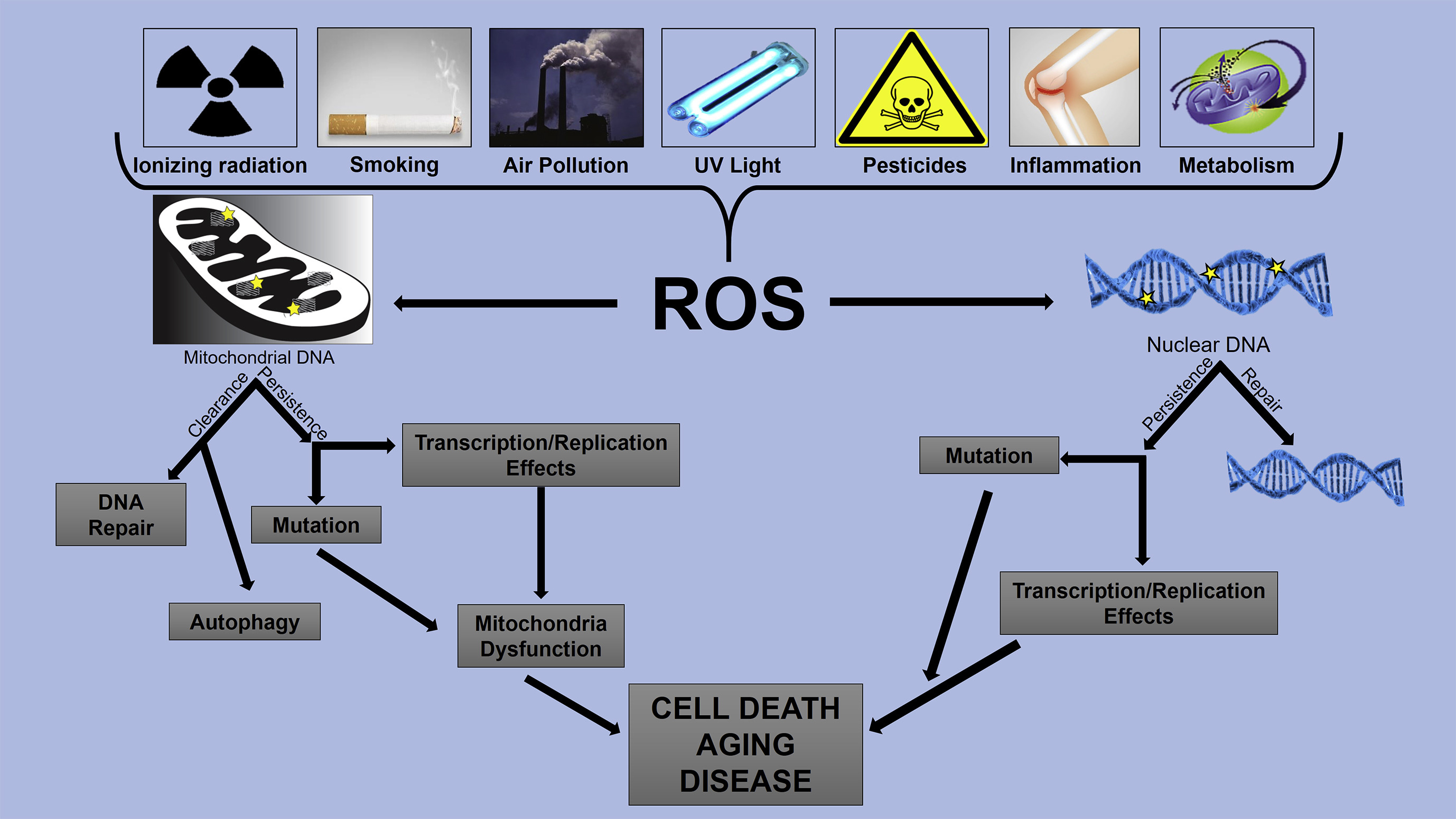
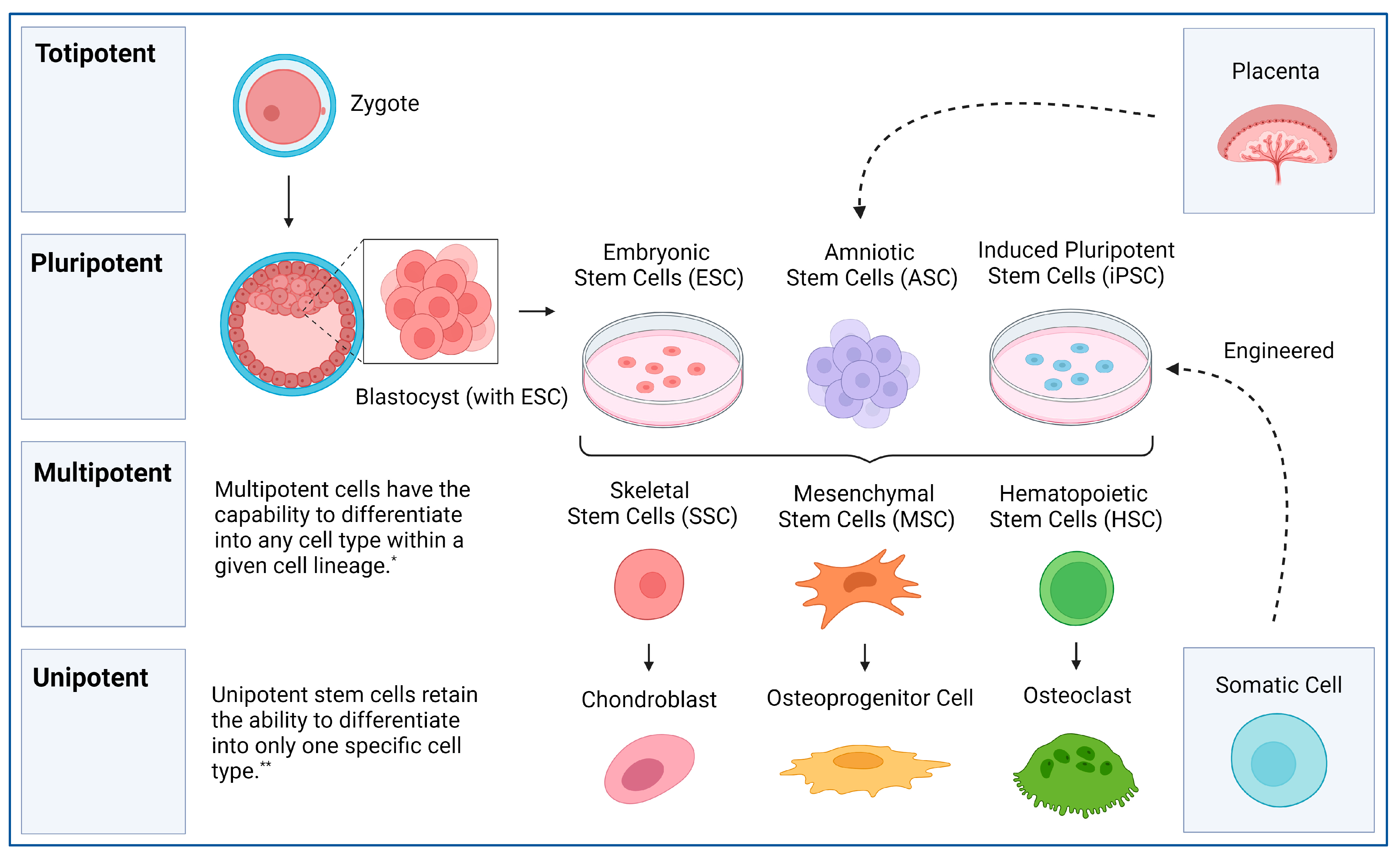
Aging happens as our cells accumulate damage over time. DNA mutations, oxidative stress (related to too many reactive oxygen species), and the shortening of telomeres (the protective caps on our chromosomes) all contribute to tissue decline. Stem cells, which can develop into different cell types, naturally diminish with age, reducing the body’s ability to repair itself.

Stem Cells as a Fountain of Youth

Scientists are investigating whether replenishing or rejuvenating stem cells could combat aging. Mesenchymal stem cells (MSCs), found in bone marrow and fat tissue, secrete growth factors that aid tissue repair. Studies suggest they could improve skin elasticity, reduce wrinkles, and even regenerate damaged organs. MSCs are already in clinical trials for treating age-related frailty and inflammation.

Another promising option is induced pluripotent stem cells (iPSCs), adult cells reprogrammed into a stem-like state. In mouse studies, researchers extended lifespan and reversed signs of aging by introducing rejuvenated cells. iPSCs could replace aged or damaged cells, rejuvenating tissues without the need for donors. In discussions, we debated whether pursuing cellular youth might create societal imbalances, benefiting the wealthy while leaving others behind.

Stem cell hierarchy from totipotent to unipotent cells, showing differentiation pathways from zygote to specialised cells. Pluripotent stem cells (ESCs, iPSCs) give rise to multipotent stem cells, which further specialise into specific cell types. Amniotic stem cells and engineered iPSCs are also depicted. 2

Stem Cell Therapies for Aging-Related Diseases

Stem cells might not just help with wrinkles – they could tackle diseases of aging. For instance, stem cell transplants are being explored for neurodegenerative conditions like Alzheimer’s and Parkinson’s. By generating healthy neurons, scientists hope to replace the lost brain cells causing these diseases.

Stem cells also show promise for cardiovascular disease. Researchers are working on generating new heart muscle cells from iPSCs, which could be transplanted into damaged hearts. Similarly, MSCs are being tested to repair cartilage in osteoarthritis patients, offering hope for those with joint pain and reduced mobility.

Rejuvenating the Skin and Immune System

Short video explaining therapeutic potential of exosomes. 3

One of the most visible signs of aging is skin deterioration. Stem cell-based treatments, like exosome therapies (using stem cell-derived vesicles filled with growth factors), aim to boost collagen production, improve skin texture, and enhance overall skin resilience.

Stem cell therapies may also rejuvenate the immune system. The thymus, which produces immune cells, shrinks with age, weakening immunity. Researchers are exploring whether stem cell injections could regenerate thymic tissue, restoring immune function and boosting longevity.

The Ethical and Practical Challenges

Despite the promise, stem cell therapies pose ethical and logistical challenges. While iPSCs bypass the controversy of embryonic stem cells, safety remains a concern. Unchecked cell growth could cause cancer, and immune responses to transplanted cells must be addressed. The technology is costly and primarily accessible through clinical trials, raising questions about equitable access – a point that sparked intense class discussions.

From a societal perspective, extended lifespans prompt complex questions: are we prepared for a world where people live to 120 or beyond? How might this affect resources and healthcare systems? These conversations made me reflect on whether the goal should be radical life extension or enhancing health span so people age with dignity and vitality.

A Glimpse Into the Future

While we’re not close to immortality, stem cells offer a promising path to healthier aging. As research progresses, therapies could shift from experimental to routine, helping people live longer, more vibrant lives. The idea of eternal youth may not be science fiction forever – with stem cells, it just might become reality.

Would you want to know what you’d look like at 150? The future of aging is unfolding, and stem cells are at the heart of the revolution.

Sources

  1. DNA damage by oxidative stress: Measurement strategies for two genomes https://ars.els-cdn.com/content/image/1-s2.0-S2468202017301341-gr1_lrg.jpg (accessed: 06/03/2025)
  2. Stem Cells and Acellular Preparations in Bone Regeneration/Fracture Healing: Current Therapies and Future Directions https://www.mdpi.com/cells/cells-13-01045/article_deploy/html/images/cells-13-01045-g001.png (accessed 07/03/2025)
  3. What is the therapeutic potential of exosomes? https://www.youtube.com/watch?v=NQeY_oIMNII&ab_channel=ScienceAnimated (accessed 07/03/2025)

ProstheTikTok – Social media as a tool for prosthetics education

For many, their only exposure to prosthetics has been from film, such as Captain Hook in Peter Pan – however, reality is far from fiction. I feel many people don’t know the truth of prosthetics and orthotics; but they’re not the mystery they seem! I myself have used an orthosis, needing a knee brace after a figure skating injury. 

With the rise of social media, it’s becoming easier than ever to share your life with others – whether that be your opinions on the latest album release, or an account of your personal experiences. One use of social media that I find fascinating is how TikTok is being used as a tool for education on prosthetics – a tool I used when wanting to learn more following the UOSM2031 lectures.


Case Study 1 – Luke Tarrant

After a motorcycle crash in Columbia, Luke was left in hospital, battling sepsis, eventually having his left leg amputated. Even with these hardships, Luke is now using his voice for good – showing every up and down on TikTok, describing the page as his ‘open journal’. 

One obstacle Luke faced was returning to a wheelchair over Christmas. After using a poorly fitting prosthetic, swelling of the residual limb left his socket unable to fit correctly. To avoid further complications, he had to take a break, making him feel like progress had been lost.

Despite the challenges, Luke remains positive – sharing the excitement he felt after being able to wear the prosthetic again. After recovery, Luke plans to get back out adventuring, hoping to finish his motorcycle journey across South America – this time with a new leg.

@luke_tarrant

So the plan is this: enjoy the next couple of weeks over Christmas and new year with some selective prosthetic wearing. Then in January, have a full month of no walking (my worst nightmare). If that doesn’t chill things out a bit, it could be back onto the operating table for some more surgery on the stump. It’s an incredibly tough spot I’m in at the moment and without the @Dorset Orthopaedic prosthetics team and specialist doctors I honestly don’t know what I would do. This too shall pass.

♬ To Build A Home – The Cinematic Orchestra

Case Study 2 – Erica the Glamputee

At 19, Erica contracted bacterial meningitis which ultimately led to sepsis. As a result of this, Erica is now a quadruple amputee. Under the name ‘Glamputee’, Erica creates informational videos on TikTok, which she hopes can be used an educational tool. 

As a quadruple amputee, Erica uses a full set of prosthetic arms and legs. She opts for ‘hook style’ prosthetic hands, as opposed to the new electronic styles. To some, this may seem confusing, but Erica explains that these electronic hands would be more restrictive for her as they can be slow and incredibly difficult to use – and expensive! 

These prosthetics make Erica more independent in her daily life, allowing her to work, care for her children and more – without prosthetics slowing her down.


Case study 3 – P&O with Mo

Mo isn’t an amputee like Luke and Erica – rather she’s a prosthetics and orthotics student at the Baylor College of Medicine. She started a TikTok page after feeling that there is a lack of understanding in what being a prosthetist or orthotist entails.

In the UK, to become a prosthetist like Mo, you first need to complete an approved degree in prosthetics and orthotics, then by law you must register with the Health and Care Professions Council. Only registered prosthetists can fit prosthetic limbs, and the devices are regulated by the Medicines and Healthcare products Regulatory Agency. 

‘Make it with Mo’ is a mini-series that Mo has created on TikTok. In each video, Mo shows the process start to finish of building the prosthetic or orthotic, with in-depth explanations about every step, something I’ve found to be incredibly informative. 

@pnowithmo

Patients can have a part in creating their prosthesis too! Laminations allow them to get creative and make their socket unique. Patients have brought their favorite tshirt designs, national flags or fun patterns they find in fabric stores to decorate their sockets. While some prefer to make sure the design can match any of their outfits, others choose to make a fashion statement. It’s a great way to help patients transition comfortably into this new lifestyle, especially if they are first time users! #prosthetics #orthotics #prostheticsandorthotics #transtibial #llp #transtibialamputation #amputation #limbloss #lowerlimbprosthesis #lamination #medicalschool #orthoticsandprosthetics #makeitwithmo #pnowithmo

♬ Hip Hop with impressive piano sound(793766) – Dusty Sky

While social media can be damaging, it can also be an incredibly useful tool for education. It is great to know there are creators out there dedicated to increasing the public knowledge of prosthetics – and I’ve loved learning from them!

What’s your favourite social media platform for educational videos, and who is your favourite prosthetics content creator?


All videos are sourced from TikTok, following the sites Terms of Service and Privacy Policy. All information on case studies is sourced from the individuals own TikTok page, as well as my own opinions.

Who really owns your body?

The future’s here! But who’s in control…

Imagine this: it’s 2080. Your heart isn’t just a meagre organ but a 3D printed wonder. You look down at your new, bionic arm, which is not just a replacement but an upgrade. With rapid advances in stem cell research, engineered tissues, prosthetics, and bionics, the lines between the human body and technology are increasingly blurring, arguably for the better.

But with these innovations comes a huge question… if a company builds part of your body, do you still own it?

Patents and Parts- Can Someone Own a Piece of You?

Oddly enough, body parts have been the centre of legal disputes before. In 1990, John Moore, a resident of Seattle, USA, was given treatment for hairy-celled leukaemia, where he was advised to undergo surgery to remove his spleen. He was also asked for permission to contribute to medical research, which he explicitly refused. Moore sued his doctor after discovering that his cells were being used for research without his permission, and resulted in a lucrative patent. The court ruled that he had no rights over his cells once they removed from his body: he lost. A summary of this interesting case study has been attached in a video below.

Fast forward to 2025: say a company patents your lab grown heart, or bionic arm; do they now own this technology that’s inside you? Various companies already limit repairs to medical devices such as pacemakers or implants, meaning you can’t legally fix them without their approval.

Cancel the Netflix- You Need to Subscribe to your Heart!

Here’s a concerning thought: what if your life changing, revolutionary implant came with a monthly fee? Indeed, this seems a dystopian concept, but not entirely far fetched. A range of pacemaker manufacturers already have restrictions on accessing software updates, and there are concerns that future medical technology such as smart prosthetics could adopt a subscription models.

A range of ethical questions are ultimately raised from this:

  • What happens if someone cannot afford their payments? Does this simply result in a loss of function of their vital organ/limb? Will this lead to death?
  • Should companies be permitted to charge for continued access to essential body parts?

A Fine Line- Treatment and Enhancement

Its almost inevitable with the pace that technology is moving, bionic limbs will eventually supersede our natural ones. Should athletes be using these in various sports? A new brain implant has the ability to boost and promote intelligence- how do we decide who has access to this… the big CEOs? Or the struggling students?

This all touches on elements of transhumanism (a video explaining this attached below), social inequalities and legal restrictions.

Conclusion: Your Body. Your Rights. But for How Long?

The idea that a company can own a part of your body might sound crazy, but we’re already heading in that direction. From patented lab-grown organs, to bionic limbs with restricted upgrades, the future of medicine is becoming entangled with corporate control, legal loopholes, and ethical issues.

And that’s an issue.

With spectacular technological advances such as bionics and prosthetics having power to transform lives we have a clear underlying risk: the commodification of our bodies. By not pushing for clear legal protections now, we’re looking at a future where vital medical technology is obstructed behind paywalls, where our own body data is exploited, and where only certain demographics such as the wealthy can partake in ‘human enhancement;’ controversial in itself.

So… who owns your body? Right now, you. But that might not be the case for much longer. Its time for society, including scientists, lawyers, and everyone in between, to ensure the future of engineered body parts is driven by human rights, not corporate profits.

Type 1 Diabetes: The fight for a cure

Imagine if your ability to eat relied on whether you could solve a handful of maths questions, lines of algebra blocking you from simply reaching over to the snack drawer and grabbing a bag of your favourite crisps. Sounds like a nightmare, right? But for 464,000 people in the UK alone, this is a version of their reality whilst living with type 1 diabetes, an autoimmune disease that prevents your body from producing insulin [1].

Insulin, a vital hormone produced by the pancreas after the ingestion of food, enables our body to transfer glucose from our blood into our cells, allowing it to be used as energy [2].

Image available at: https://gluroo.com/blog/diabetes-101/insulin-faqs-type-1-diabetes/ (accessed: 09/03/25)

Those with type 1 diabetes have a pancreas that is unable to produce enough insulin to promote this transfer causing their levels of blood glucose to slowly increase with each meal, the sugars predominantly being expelled only as waste via frequent urination. As a result, insulin must be supplemented through injection, quantities being meticulously calculated dependent on what is being eaten and when. Whilst with years of practice calculating the amount of insulin you need can become almost second nature, many with this disease long for a day where they can eat freely, not having to constantly monitor their blood sugar levels and symptoms.

A short video explaining some key aspects of type 1 diabetes – closed captions are available!

Current Technologies

Whilst technology currently exists which aims to make the lives of those with diabetes easier, nothing works quite as well as a pancreas. For example, DexCom is a company which produces a small sensor which allows for the continuous monitoring of blood sugar levels, a connecting app receiving and displaying the statistics on a regular basis. Having an error of only 8.2%, this sensor would appear to be amazing in almost any other application, but when it comes to someone’s health you can never be accurate enough [3].

This video follows a young woman called Jordan and how DexCom allowed her to regain control of her life and love for sport (closed captions available)

With pancreas transplants carrying the same risks as any other major surgery, the possibility of rejection urging healthcare professionals to encourage the majority to manage their condition through diet and medication, most are left with these sensors as their most advanced form of care. Therefore, although some aspects of life with type 1 diabetes can be made more “convenient”, the fight for an accessible cure is still ongoing.

Stem Cells and Ethics

Excitingly, it has been reported that a 25-year-old woman with type 1 diabetes has undergone a stem cell transplant enabling her pancreas to begin producing insulin again, it continuing to do so even a year post transplant – more about this encouraging story can be read here! Whilst this seems promising, the procedure does come with risks. It is believed that a form of immunosuppression is required in order to prevent complications, making the patient more vulnerable to other illnesses and infections.

Furthermore, there is currently a large volume of debate over the ethics of using stem cells in treatments. Embryonic stem cells, those that are typically used in such treatments, can only be obtained through the destruction of a human embryo, a process that breeds concern surrounding the sanctity and rights of early human life [4].

Image available at: https://bioinformant.com/what-are-stem-cells/ (accessed: 09/03/25)

Whilst many believe using a potential human life in this manner is unethical, I can’t help but wonder if denying somebody a better quality of life that could be so easily provided is just as cruel. How would you feel if a cure to something that controlled your daily life was just out of reach?

References

[1]Diabetes UK, “How Many People in the UK Have diabetes?,” Diabetes UK, 2024. https://www.diabetes.org.uk/about-us/about-the-charity/our-strategy/statistics

[2] NHS , “What is type 1 diabetes?,” nhs.uk, Nov. 31, 2024. https://www.nhs.uk/conditions/type-1-diabetes/what-is-type-1-diabetes/

[3]S. K. Garg et al., “Accuracy and Safety of Dexcom G7 Continuous Glucose Monitoring in Adults with Diabetes,” Diabetes Technology & Therapeutics, vol. 24, no. 6, pp. 373–380, Jun. 2022, doi: https://doi.org/10.1089/dia.2022.0011.

[4] L. A. Cona, “Stem Cell Research Controversy: A Deep Dive (2023),” www.dvcstem.com, Sep. 14, 2023. https://www.dvcstem.com/post/stem-cell-research-controversy

Cloning: Simply Science Fiction?

The idea that an individual could be identically replicated has captivated popular culture for decades. A clone is defined in biology as an organism or cell, produced asexually from one ancestor, to which they are genetically identical. Appearing in the likes of the Star Wars franchise, Oblivion, and more recently Mickey-17 released just this month, clones in science fiction films are often portrayed in a way that challenges this definition. When reproductive cloning and Dolly the sheep were introduced in Nick Evans’ lecture on stem cells, I asked myself the following questions:

  • To what extent does cloning exist?
  • Why haven’t we cloned humans?
  • What are the applications of cloning?

Animal cloning – Dolly the sheep

On the 5th of July, 1996, the first mammal cloned from an adult somatic cell was born – Dolly the Finn-Dorset sheep. The scientific advancement captured the attention of the media at the time, and continues to do so today (see links on right). The procedure involved somatic cell nuclear transfer, which is a type of reproductive cloning. The diagram below summarises the method.

Image from Encyclopædia Britannica, Inc.

Dolly essentially had three mothers, with the DNA from the egg cell of a Scottish Blackface sheep being replaced by the DNA from a Finn-Dorset sheep. The resulting hybrid cells were placed in the uterus of another Scottish Blackface, and astonishingly, Dolly was born with identical genes to the nucleus donor sheep. In theory, the exact same procedure could be completed with humans. It should be noted that it took 227 attempts to produce one sheep clone. Even today, mammalian cloning is highly inefficient.


Ethical Considerations

Following the birth of Dolly, the United Nations Educational, Scientific, and Cultural Organisation (UNESCO) immediately banned cloning. It goes without saying that cloning humans would raise innumerable ethical issues. Below are the most notable (click on the arrows for more information):

Eugenics

An early ethical consideration, following John Lederberg advocating for cloning in 1966. He saw it as an appropriate means to improve the human race – cloning certain individuals for their desirable traits. Many see this is a ‘new form of eugenics’, which is considered immoral by most, due to questions of equality and coercion.

Danger

Utilitarianism focuses on the direct outcome, raising the concern that cloning human beings by nuclear transfer is too dangerous. Too many embryos in animal cloning experiments expire or result in miscarriage.

Individuality

Many believe that cloning violates our dignity, uses people as means, affronts our uniqueness, and threatens our humanity.

Embryonic research

To even research human cloning, a huge amount of human donated eggs would have to be acquired by scientists. The nature of the nuclear transfer method, with implantation would also require the research to surpass the 14-day limit of embryo experimentation, raising questions on identity and the value of human life.


Therapeutic Cloning

Therapeutic cloning is a practical method which in my opinion avoids the ethical issues raised above. It also involves nuclear transfer, but the cells are never implanted. Instead, the embryo is cloned to produce stem cells. Since these would be genetically identical to the donor, these stem cells are less likely to be rejected by the patient.

Image from Mitalipov Lab/OHSU

Conclusion

It seems to me that human cloning is inefficient, immoral, and mostly pointless. The replicas we see in science fiction don’t reflect biology, and with therapeutic cloning, there is no need to clone entire humans for treatment.

“Your scientists were so preoccupied with whether or not they could, they didn’t stop to think if they should.”

Ian Malcolm, Jurassic Park (1993)

3D Bioprinting

Press a button, print an organ. Perhaps the idea is not as Sci-Fi as it sounds. 3D bioprinting is an advancing field that uses layer-by-layer positioning of cells and biomaterials to produce living scaffolds for a variety of uses.

Depending on the qualities needed in the final product, techniques and materials vary. All start with a digital 3D model produced using computer aided design (CAD) software. One powerful feature of this is that CT and MRI scan data from patient can be used to tailor, for an example a tracheal graft, to personalised dimensions.

Next, the structure can be bioprinted using a bioink. Things to consider when choosing a bioink are the cells, biomaterial and growth factors included. Biomaterials such as alginate and polyvinyl-alcohol provide a structural scaffold for new cells to grow on. The ideal biomaterial must be biocompatible (not trigger an immune response) and biodegrade at the same rate as the new tissue grows.                                     

Cells and growth factors goes hand in hand, to print a bone graft you would use mesenchymal stem cells and a corresponding growth factor or chemical that encourages the pathway of differentiation looked for. The bioink used can dictate the printing method. Three popular approaches include inkjet-based, laser assisted and extrusion-based bioprinting. Each has their own set of opportunities and challenges.

Inkjet bioprinting uses the bioink droplets deposited by a piezoelectric actuator. Laser-assisted uses an a donor layer than absorbs energy from laser stimulation with a bioink layer underneath it and a collecting layer to form the tissue constructs. Extrusion uses mechanical force to deposit a continuous cylindrical stream of bioink.
Mechanisms of 3D bioprinting: Adapted from an image by Loai et al, licenced under Creative Commons Attribution 4.0 International License from https://doi.org/10.20900/rmf20190004

Droplets of bioink, generated either through heating or a piezoelectric effect, are deposited on the substrate. High throughput, high resolution, low expense and strong cell viability count in favour of this technique. The key drawback is the need for low viscosity bioink to pass through the fine nozzle which results in low structural quality.

If a strong structure is a priority then extrusion printing comes to the fore. It relies on pneumatic or mechanical force to print uninterrupted lines of a far more viscous bioink. However, the increased shear stress involved reduces cell viability. High viscosity bioinks tends to result in lower resolution and the process is much slower.

This uses a laser induced forward transfer technique (LIFT) which means that a laser is used to force material from a thin donor layer of bioink onto the substrate being printed onto. Advantages of this include high cell viability as there is no direct contact between the bioink and the dispenser so no stress forces and that the technique is compatible with a wide range of bioink of varying viscosities. Unfortunately, the complexity of this method results in high equipment cost.

Potential Uses

  • Organ replacement
  • Tissue grafts such as skin, bone muscle and cartilage
  • Organs on a chip – used for drug and vaccination screening
  • Drug delivery scaffolds
  • Building disease models

Printing organs for those currently awaiting a donor remains science fiction for now but great progress is being made towards that goal. However, even this is just the start as ethical considerations need to be made.

Integral to the whole process are stem cells and their source. The ethics of stem cells use are hotly debated but the alternative, induced pluripotent stem cells, are associated with more unpredictable behaviour.

Safety and quality control of bioprinted organs need to be considered, and this is made harder personalisation of treatment which reduces replicability of results. Arguably, for a terminally ill patient the benefits of an untested treatment outweigh the risk, but can this then be applied to non-life-threatening cases?

A New You! Stem Cells in Regenerative Medicine

Can the body heal itself? The power of stem cells in a new era of healing

Stem Cells are cells that can self-renew and differentiate, this is crucial for the development of an organism as well as repair after injury (Mayo Clinic, no date). As a cornerstone of regenerative medicine, they offer innovative new solutions to treating diseases with limited treatment options such as restoring damaged organs and tissues (Wang et al. 2024).

Understanding Stem Cells

It is important to understand the different types of stem cell

  • Multipotent: The ability to differentiate into more than one cell type in the body
  • Pluripotent: The ability to differentiate into all of the various cell types in the body

Pluripotent and multipotent stem cells originate from different sources. Pluripotent stem cells are derived from early-stage human embryos, these are capable of dividing without differentiation and can develop into the primary three germ layers. In comparison, adult stem cells can only differentiate into the cell type of the tissue that they are found (Mayo Clinic, 2025). Researchers have developed an induced pluripotent stem cell, similar to embryonic stem cells, but, formed by transferring embryonic genes to a somatic cell (Mayo Clinic, 2025).

What is Regenerative Medicine

Regenerative medicine is a branch of medicine that focuses on healing or replacing organs and tissues damaged by factors such as disease or trauma. ‘Healing’ is achieved by replacing missing tissue structurally or functionally using stem cells such as mesenchymal stem cells induced pluripotent. This is achieved through materials as well as de novo-generated cells. It is also possible to leverage the body’s inate healing response, however humans lack regenerative capacity (Mao and Mooney, 2015). Regenerative medicine has the potential to treat neurodegenerative diseases as well as heart failure, for example.

Stem Cells in Regenerative Medicine

Adult stem cells such as mesenchymal and induced pluripotent stem cells (iPSCs) , exhibit multilineage differentiation capacities as well as immunomodulatory properties (Li, Luanpitpong, and Kheolamai, 2022). There have been successful treatments using these stem cells for bone and cartilage regeneration as well as spinal cord injuries and diabetes (Hoang et al., 2022). allowing them to differentiate into required tissues and treat injuries, inflammation, and age-related disorders through regeneration of muscle, cartilage, and muscle regeneration. Human pluripotent stem cells (hPSCs), embryonic stem cells (ESCs), and (iPSCs) can differentiate into any cell type so have a very high potential (Wang et al., 2024).

Challenges Associated

However, there remain risks associated with stem cell use such as a heightened risk of tumor formation, and immune rejection, and it’s not guaranteed that the cells will survive post-translation.

The treatments with the most therapeutic potential use embryonic stem cells, due to their pluripotency. However, the obtainment of ESCs is controversial due to the destruction of human embryos, many people object to religious and ethic principles (Margiana et al., 2022). Stem cell therapies also have a high cost meaning availability is restricted to wealthier patients (Hoang et al., 2022).

Conclusion

The advancement of regenerative medicine presents exciting opportunities such as within genetic modification, combining stem cells with drug delivery as well as biomaterial scaffolds (Li, Luanpitpong, and Kheolamai, 2022). However, currently stem cells remain at the forefront for conditions such as cardiovascular disease and organ regeneration. However, further research is needed to combat concerns such as immune rejection and tumor risks. For this treatment to become mainstream steps will have to be taken to address high costs and accessibility issues as well as the need for a global regulatory body to monitor its use.

Bibliography

Hoang, Duc M., Phuong T. Pham, Trung Q. Bach, Anh T. L. Ngo, Quyen T. Nguyen, Trang T. K. Phan, Giang H. Nguyen, et al. ‘Stem Cell-Based Therapy for Human Diseases’. Signal Transduction and Targeted Therapy 7, no. 1 (6 August 2022): 1–41. https://doi.org/10.1038/s41392-022-01134-4.

Li, Jingting, Sudjit Luanpitpong, and Pakpoom Kheolamai. ‘Editorial: Adult Stem Cells for Regenerative Medicine: From Cell Fate to Clinical Applications’. Frontiers in Cell and Developmental Biology 10 (31 October 2022). https://doi.org/10.3389/fcell.2022.1069665.

Mao, Angelo S., and David J. Mooney. ‘Regenerative Medicine: Current Therapies and Future Directions’. Proceedings of the National Academy of Sciences 112, no. 47 (24 November 2015): 14452–59. https://doi.org/10.1073/pnas.1508520112.

Margiana, Ria, Alexander Markov, Angelina O. Zekiy, Mohammed Ubaid Hamza, Khalid A. Al-Dabbagh, Sura Hasan Al-Zubaidi, Noora M. Hameed, et al. ‘Clinical Application of Mesenchymal Stem Cell in Regenerative Medicine: A Narrative Review’. Stem Cell Research & Therapy 13, no. 1 (28 July 2022): 366. https://doi.org/10.1186/s13287-022-03054-0.

Mayo Clinic. ‘Answers to Your Questions about Stem Cell Research’. Accessed 5 March 2025. https://www.mayoclinic.org/tests-procedures/bone-marrow-transplant/in-depth/stem-cells/art-20048117.

Mayo Clinic. ‘Answers to Your Questions about Stem Cell Research’. Accessed 5 March 2025. https://www.mayoclinic.org/tests-procedures/bone-marrow-transplant/in-depth/stem-cells/art-20048117.

Wang, Jipeng, Gang Deng, Shuyi Wang, Shuang Li, Peng Song, Kun Lin, Xiaoxiang Xu, and Zuhong He. ‘Enhancing Regenerative Medicine: The Crucial Role of Stem Cell Therapy’. Frontiers in Neuroscience 18 (8 February 2024). https://doi.org/10.3389/fnins.2024.1269577.

Petri Dish to Patient : How 3D Bioprinting is Changing Transplants

“Why can’t we just print more organs?” Overcoming the organ shortage crisis with Bioengineering

Globally, the demand for organ transplants exceeds supply. In the United States as of 2021 116,566 patients were waiting for an organ transplant, 6 564 patients died waiting for an organ. Despite this, only 41 354 transplants were performed (Kupiec-Weglinski 2022). 3D bioprinting is an emerging technology that offers a promising solution by creating bioengineered organs and tissues, reducing patients left waiting for life-saving treatments and reliance on human donors (Bose, 2023)

3D bioprinting is an innovative technology that involves printing layer by layer, combining biomaterials, bio-inks, and living cells (Panja et al., 2022). This provides an opportunity to engineer human tissues and organs for medical use such as :

  • Drug testing on human models (more reliable and reduces demand for animal models)
  • Custom implants
  • Reducing the waitlist for replacement human organs

How It Works

There are 4 main bioprinting techniques

Technique How it Works Pros Cons
Extrusion Bioprinting Uses mechanical force to push bio-ink through a nozzle Good for large tissues
Can use high-viscosity bio-inks
Can cause damage to cells from pressure
Laser-Assisted Bioprinting Uses lasers to deposit biomaterials High precision
No nozzles needed
Expensive, Slow
Stereolithography (SLA)Uses UV light to harden layers of biomaterials High resolution, Fast Limited material options
Inkjet Printing Droplet-basedFast, Cost effective Low precision
Can only use low-viscosity bio-inks

(Panja et al., 2022)

Applications

Recently there have been significant breakthroughs in replicating complex human organs especially hollow organs such as the lungs, heart, and digestive system. Despite the immense progress, technical challenges still persist.

Bioprinting Lungs 

  • Lung diseases such as COPD and COVID-19 have increased demand for lung transplants. The focus remains on the airways and alveoli.  Advancements have been made in the development of artificial alveolar models using inkjet printing, hydrogels, and synthetic polymers.  Despite this, these have not been applied to organ replacement yet (Panja et al., 2022). Achieving the highly complex structure and function of alveoli is still a significant hurdle. 

3D Printed Heart Tissue 

  •  A heart has been bioprinted including blood vessels. This is crucial when developing the functionality of replacement organs. This was achieved using a technology called Coaxial Sacridicial Writing in Functional Tissue. This adds layers of real blood vessels, blood supply is essential for sustaining all bioprinted organs (Brownell, L. 2024).

Digestive System 

  • Researchers have also seen developments in the bioprinting of the digestive system, including stomach, intestines and bile ducts. There is enormous potential for this to revolutionize drug testing and reduce reliance on animal models. However, barriers remain due to the inability to replicate the mechanical processes of the digestive system such as peristalsis(Panja et al., 2022. 


Challenges and Ethical Concerns

  • Cost: Bioprinting is an expensive technology, with significant costs from research, development, and clinical trials. As well as the extensive laboratory equipment, this means that the early technology is likely to be limited to the wealthy (Bose, P., 2023).
  • Tissue Vascularisation: functional blood vessels are imperative for delivering oxygen and nutrients, and keeping tissues alive. Functional blood vessels are challenging due to their microscopic size and complexity (Panja et al., 2022).
  • Regulation and Safety: international regulations are necessary to ensure safety, efficacy and ethics have been taken into consideration (Brownell, L. 2024).

Conclusion

3D printing has the potential to make fully functional, transplantable organs a reality. There needs to be a focus on personalization to reduce the risk of elimination. However, vascularisation will remain a significant barrier to applicability. However, the potential to remove donor waitlists and animal models.

Could we be entering an era of on-demand organ transplants?

Bibliography

Bose, P. (2023). Bioprinting Organs: A Look into the Future of Transplantation. News-Medical. Available at: https://www.news-medical.net/health/Bioprinting-Organs-A-Look-into-the-Future-of-Transplantation.aspx [04/03/25].

Brownell, L. (2024). 3D-printed blood vessels bring artificial organs closer to reality. Harvard John A. Paulson School of Engineering and Applied Sciences. Available at: https://seas.harvard.edu/news/2024/08/3d-printed-blood-vessels-bring-artificial-organs-closer-reality [04/03/25].

Panja, N., Maji, S., Choudhuri, S., Ali, K. A., & Hossain, C. M. (2022). 3D Bioprinting of Human Hollow Organs. AAPS PharmSciTech, 23(5), p.139. Available at: https://doi.org/10.1208/s12249-022-02279-9 [04/03/25].

Kupiec-Weglinski, Jerzy W. ‘Grand Challenges in Organ Transplantation’. Frontiers in Transplantation 1 (6 May 2022). https://doi.org/10.3389/frtra.2022.897679. [04/03/25]

Knee replacements: Cost for Mobility

“We don’t appreciate what we have until it’s gone.” This phrase resonated deeply with me during the lecture on joint replacements. We often take for granted the ability to move and stand without pain—until one day, we can’t. Millions of lives have been transformed by the invention of knee replacement, restoring people’s movement but also their independence. But what are the challenges of these replacements and what does their future look like?

Evolution of knee replacements

The idea of replacing damaged joints isn’t new and has been around for hundreds of years where ancient civilisations experimented with rudimentary prosthetics. Modern knee replacement surgery, or total knee arthroplasty (TKA) however, only emerged in the mid-20th century. Advances in materials, surgical precision, and rehabilitation have meant that patients today can regain almost all lost function. Whilst these improvements have been greatly beneficial, there are still risks and limitations involved.

image: Fixed vs mobile bearing of knee replacements https://link.springer.com/chapter/10.1007/978-981-16-8591-0_3

More Than Just a Mechanical Problem?

Engineering is at the core of a knee replacement where surgeons remove damaged bone and replace it with metal and synthetic components that are specifically designed to mimic the joint’s natural function. The real challenge however, is in how the body allows the artificial joint to work within our bodies, where ligaments, tendons, and muscles must adjust to the artificial joint, and imbalances in force distribution can lead to complications such as implant loosening. The risk of rejection is also prevalent as the body may recognise the implant as foreign, triggering low-grade inflammation that can affect the implant’s longevity.

As with any surgery there is the emotional and psychological side. How does it feel having to relearn simple movements such as walking? Regaining autonomy can be life-changing, but rehabilitation can be long and demanding.

Challenges and Ethical Considerations

Despite its success and widespread use, it still has its limitations.

  • Longevity of implants: A knee prosthesis only lasts 15–20 years and so younger patients may require multiple complex surgeries.
  • Access to prosthetics: Not everyone can afford a knee replacement. Is it right that orthopaedic treatments are more readily available to those with financial means?
  • Surgical risks: Infections and rejection are still major risks.

In a recent BBC article from last year, Heather considered herself “lucky” to have had her left knee replaced after two years of pain. This article highlights a wider systemic issue in healthcare-availability. The question remains as how can we solve this?

This shows that there is still a long way to go when talking about this type of technology and highlights the need for continuous innovation in this area.

Future of Knee Replacements: Can We Do Any Better?

With recent advancements in areas such as 3D printing and robotic-assisted surgery, the future of knee replacements is still promising. One day, could we use cartilage grown in a lab to produce personalised implants? And could AI-driven rehabilitation programs increase patients’ recovery outcomes?

This lecture changed the way I think about joint replacements as a field that intersects engineering, biology, and ethics. It made me think about the the privilege of mobility and the importance of continuous innovation in medical technology.

Bioengineering with Bubbles!

After an insightful trip to the hospital for the Stem Cell and Regenerative Medicine Masterclass, I discovered a new branch of biomedical engineering that I’d never considered: using bubbles in medicine! Now I know what you’re thinking, but these aren’t bubbles that you’d make from soapy water and by waving a wand around vigorously in the air. Instead, these bubbles are microscopic, made from a monolayer of phospholipids that self aggregate to form a sphere with a gas core. PhD student Aya Ben Issa demonstrated how to create these bubbles, alongside explaining some potential uses, including her own research focus, using them to break up biofilms. This got me thinking, what other uses for microbubbles are there?

Microbubbles for Imaging

Primarily, microbubbles are used for contrast enhanced ultrasound in medical imaging. Sounds like a mouthful, but in reality, it essentially improves the imaging quality by creating a better distinction between the tissues and the injected microbubbles. I’ve included a video so you can have a look for yourselves:

Microbubbles for drug delivery

When the idea of microbubbles was first introduced, my mind initially went to using them for delivering drugs. And why not? These micro compartments seemed like the ideal packaging for doing exactly that. With further research into the topic, I discovered that I wasn’t the only one who thought the same. Our current module lead, Professor Nicholas Evans has published an article on using them to deliver oxygen to tissues. This has shown promise for bone cancer treatments by reducing metastasis. This supports my initial thought; that things can be delivered within the bubble. But what about on them? Well, once again, studies have indicated that insertion of drugs into the microbubble shell in addition to attaching site-specific ligands have been show to allow targeted delivery of drugs to areas where pathologies have occurred.

When do Ultrasound and Microbubbles Go Hand in Hand?

So, you’ve put your drugs within your microbubbles and injected them into the patient. They’ve travelled to the target site. Now what? You need to actually get your drugs out of the bubble and into the tissue. This is where ultrasound comes in. Ultrasound is able to create oscillations and compressions within the shell of the microbubble, causing a large dose of drug to be released. In addition to the microtubules being the delivery agents of the drugs, they can also be used as mechanical agents. The ultrasound once again causes oscillations, this causes transient, miniscule disruptions in the plasma membranes of the cell, allowing drugs to once again enter the cell. This process is known as sonoporation.

Examples of Uses

In summary of the topics I have suggested above, the image below highlights some of these applications, from drug delivery to sonoporation, as well as some other potential applications.

What do I think?

In my opinion, the application of microbubbles have great potential. Of course, further studies will have to be done to fully understand the extent to which we can use this technology. There are still limitations when it comes to the therapeutic uses of these microbubbles in addition to some novel ideas such as Aya’s application of them to disrupting biofilms. What do you think? Can you think of any other uses of these microbubbles?

Want some more info?

Use the hyperlinks in this article for a more in-depth understanding of the topic I have discussed today! 🙂