The University of Southampton

Human-Animal Hybrid Brains: A Dr. Frankenstein Dilemma

Introduction:

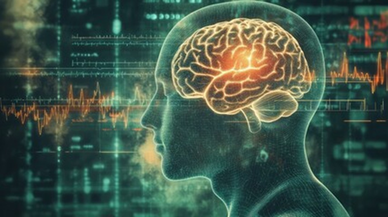
While studying for a debate regarding the use of embryonic stem cells (ESCs) in research, I stumbled across articles about organoids. I remembered them being mentioned in one of our earlier lectures, therefore these articles peaked my interest. So I dug deeper. What interested me was the slightly morbid idea of growing a human brain and what that means. This digging led to another branch of this scientific, dystopian tree: brain chimeras. In this blog, I will be discussing why brain organoids and chimeras are made, what they are used for and what ethical issues rise.

Are we as scientists, justifying the use of these chimeras the same way Dr. Frankenstein justified the creation of his monster, selfishly in pursuit of greater knowledge despite the pain and suffering this may create?

What are they and how are they made?

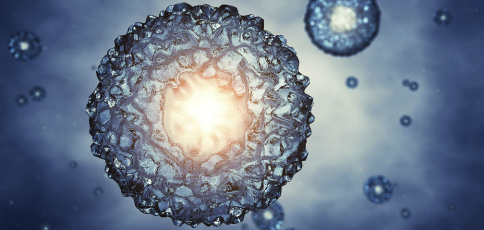
Organoids are 3D, self-organised tissue cultures made from stem cells. They are made by using iPSCs (induced pluripotent stem cells) which are made from adult somatic cells. This is done by reprogramming these somatic cells using transcription factors to make the cell pluripotent similar to ESCs.

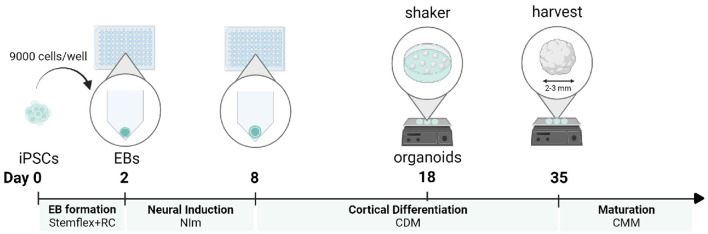
To grow these organoids the iPSCs are cultured to form an embryoid body (EB) which mimics the features of an early stage embryo. This is exposed to different mediums to encourage differentiation into brain cells then grown in a growth medium.

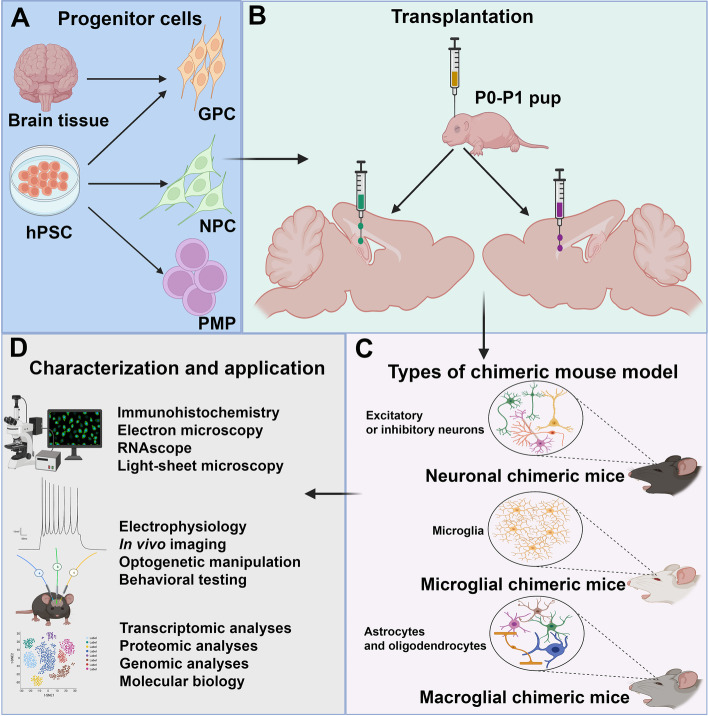
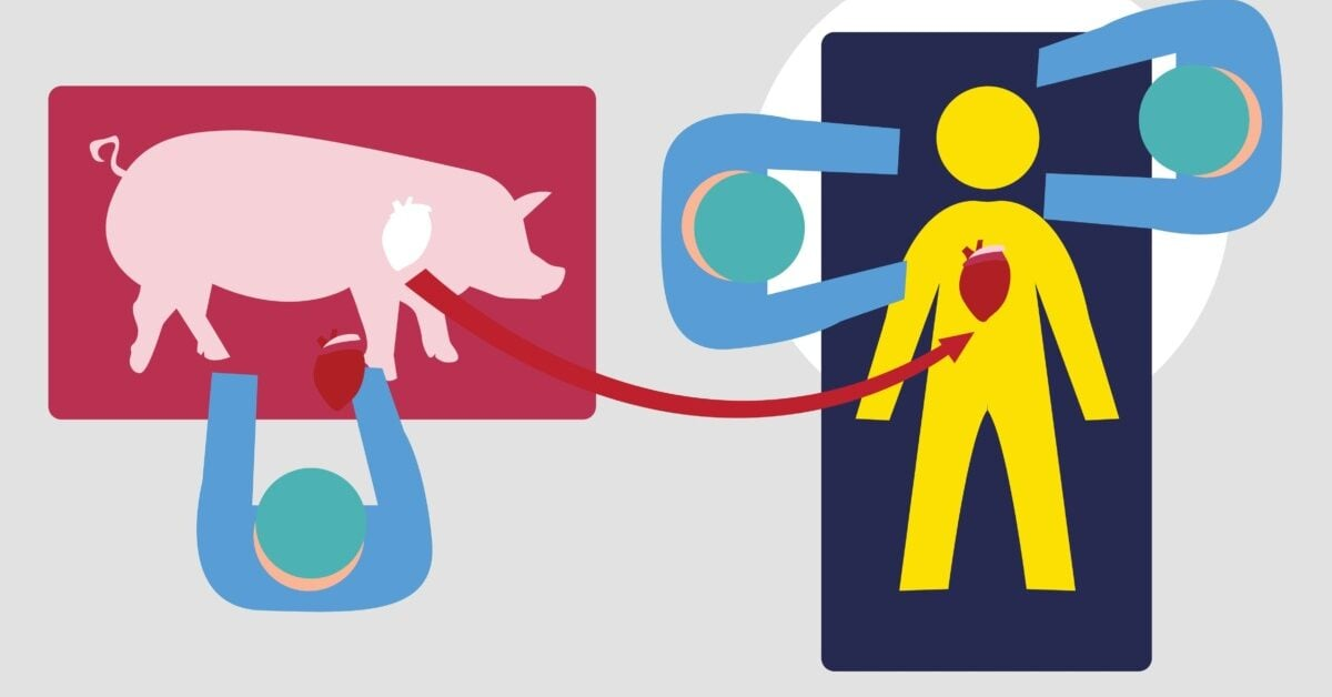
Organoids can be made with human stem cells and animal stem cells. This results in brain Chimeras which are made by engrafting human foetal tissue/iPSCs into a neonatal animal (e.g. mouse) brain.

Why are they important?

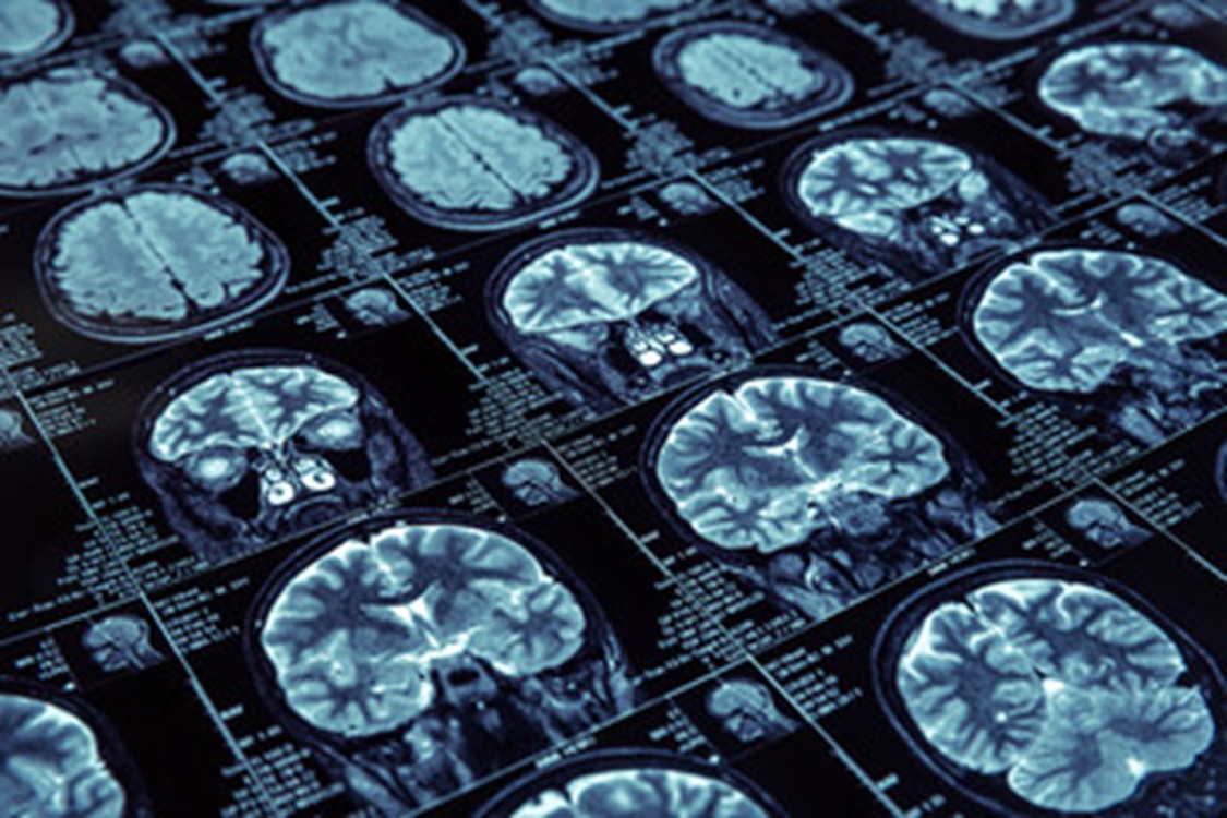
We all know someone who is battling with a neurological condition. Many of these conditions are debilitating, ruin lives and have high mortality rates. This highlights the significance of this research.

One in two of us will suffer with dementia in our lifetime. Therefore, research into neurodegenerative disease is more important than ever before.

Although these organoids and human-animal brain chimeras are relatively new there has been a tremendous volume of studies which use this technology. They can offer important insight into the progression of neurodegenerative disease leading to new treatments. However, one could argue that these ‘brains’ are still fundamentally different and less complex than a real human brain as most animals are biologically very different and display different symptoms of neurological problems.

The issue

The main debate lies with the idea that human brain organoids or human-animal brain chimeras could be capable of intelligent thought. This raises many concerns as currently organoids hold no legal or moral rights within science. Are these chimeras thinking the same as a human may think? Are they self aware? More complex than say an ordinary mouse? And why should this matter?

There are significant regulations when using hESCs for research, such as the 14 day rule, however, organoids and human-animal chimeras are not regulated the same way, it does not even require a license as long as it does not use hESCs. Why is this?

These brain organoids and chimeras have the same moral status as many animals that are used in research. Should this be the case?

There is no right answer here as you cannot possibly know what a chimera or brain organoid could be thinking.

Conclusions and Reflections

Prior to researching this topic I did not appreciate the complexity and possible future of these models, I had a similar initial reaction to using any animal model; that it is a cruel and disturbing idea. However, animal models and hESCs have contributed greatly to the world of science, without them many treatments (HIV medication, cancer medications, vaccines) would not have been developed. This shows how important in vivo research is and these organoid/chimeric models may just add to the possibilities.

Contrary to this, I was surprised to find that making an organoid does not require any sort of formal license. I believe organoids should be treated with the same moral significance as hESCs as there is a grey area regarding their consciousness.

In conclusion, organoids and chimeric models raise many ethical and thought provoking questions, however, they offer invaluable research opportunities which could help many people, although they should be treated with more moral significance similar to that of hESCs.

References:

CAPPS B. Do Chimeras Have Minds?: The Ethics of Clinical Research on a Human–Animal Brain Model. Cambridge Quarterly of Healthcare Ethics. 2017;26(4):577-591. doi:10.1017/S0963180117000093

Grenier, K., Kao, J. & Diamandis, P. Three-dimensional modeling of human neurodegeneration: brain organoids coming of age. Mol Psychiatry 25, 254–274 (2020). https://doi.org/10.1038/s41380-019-0500-7

Kwisda, K., White, L. & Hßbner, D. Ethical arguments concerning human-animal chimera research: a systematic review. BMC Med Ethics 21, 24 (2020). https://doi.org/10.1186/s12910-020-00465-7

Eigenhuis KN, Somsen HB, van der Kroeg M, Smeenk H, Korporaal AL, Kushner SA, de Vrij FMS, van den Berg DLC. A simplified protocol for the generation of cortical brain organoids. Front Cell Neurosci. 2023 Apr 4;17:1114420. doi: 10.3389/fncel.2023.1114420. PMID: 37082206; PMCID: PMC10110973.

Organ-on-Chip: A Suitable Replacement for Animal Testing?

What if your doctor could test different treatments on a miniature replica of your organs before prescribing the perfect medication for you? This futuristic idea quickly becomes a reality with organ-on-chip (OoC) technology.

I first encountered OoCs when I was assigned my dissertation topic. Before that, I had never even heard of them. But as I delved deeper, the potential applications seemed limitless, from revolutionizing drug development to unexpected uses like measuring water pollution by simulating how a fish gill interacts with its environment (the focus of my research).

What are Organ-on-Chip Devices?

An organ-on-chip is a microfluidic device constructed to mimic the structure, microenvironment, and physiological functions of organs. These devices use microfluidic channels (small tubes) to transport essential nutrients, drugs, or toxins to a culture of living cells while removing any waste products. They vary in design depending on the organ they are simulating, offering a range of parameters like chemical gradients, mechanical shear, and tissue-tissue interactions making them ideal for drug screening and toxicity testing.

The video from Wyss Institute further illustrates how one of these innovative lung-on-chip devices works and its potential in the pharmaceutical field.

Source: Introduction to Organs-on-a-Chip

Why are Organ-on-Chip Devices Beneficial?

  • Traditional 2D cell cultures cannot accurately reflect the structure, function, and mechanical behaviour of living tissue or its highly complex interactions with the rest of the body. OoCs bridge this gap by recreating key physiological conditions in a controlled microenvironment, offering better drug testing results than conventional methods.
  • Developing a new drug is notoriously slow and expensive – it can take up to 10 years and cost more than $3 billion (that’s a lot of money!). This is mainly due to the reliance on animal testing before human trials. However, animal models have a high failure rate as their biological responses often do not always match human reactions. This leads to ineffective or toxic drugs progressing through clinical trials, while potentially effective compounds never moving to market.
    OoCs address this issue by using human cells, making them more predictive of actual drug effects in people whilst being faster, cheaper, and requiring fewer resources. Allowing earlier detection of any harmful side effects earlier in development preventing costly late-stage failures.
  • Many people including myself consider OoCs a more ethical alternative to animal testing. By providing a more accurate alternative while minimizing reliance on animal testing.

Challenges of Organ-on-Chip Technology

  • The current manufacture of OoCs is often performed in research labs requiring specialized fabrication techniques. Moving to mass production while maintaining its precision poses a significant challenge.
  • Most OoCs use polydimethylsiloxane (PDMS) due to its biocompatibility, flexibility, and transparency properties to form microfluidic channels. However, PDMS absorbs small drug molecules, potentially affecting results. Finding alternative materials that do not interfere with drug testing is needed to improve accuracy.

Figure below providing further details on the fabrication process of an Organ on chip:

  • OoCs are not perfect, they are simplified models of organs replicating only the key functions of the organ failing to capture the whole function and complex role within a human body.
  • The use of human cells for OoCs still raises ethical questions, particularly regarding the sourcing of cells how will they be obtained are the doners consenting? OoC technology isn’t cheap limiting its benefits and availability across the world.
  • There is a lengthy process before OoCs can be considered a suitable alternative to animal testing. This is governed by regulatory bodies like the FDA and EMA which require extensive validation.
  • Long-term maintenance of cell cultures in OoC devices presents its own challenges, including ensuring a steady supply of nutrients, efficient removal of waste products, and maintaining stable microenvironmental factors such as pH and oxygen levels. Additionally, cellular senescence – the gradual loss of cells’ ability to divide and function further complicates its prolonged use.

The Future of OoCs Technology

OoC technology is rapidly advancing, with models already developed for organs including brain, gut, lung, heart, and more. However, there is a universal goal towards producing a human-on-chip. A human-on-chip integrates multiple OoCs into a single system, allowing simulation of organ-organ interactions within the body. This innovation would have applications in a multitude of areas:

  • Personalized medicine: using a patient’s own miniaturised organ system to test various drugs to identify the most effective treatment.
  • Drug development: providing a more effective approach by mimicking and tracking how the drugs travel through the entire body.
  • Disease modelling: demonstrating how complex diseases like cancer or diabetes impact various organs over time, accelerating research and the discovery of effective treatments and cures.

As I mentioned earlier, the potential of OoC technology is limitless and fascinating. The concept of a human-on-chip functioning as a “mini-me” to monitor health could revolutionise how we understand and maintain personal wellness. Imagine real-time updates about your body’s needs like nutrient deficiencies or predicting health issues before symptoms even appear.

While challenges remain, I am optimistic about their future and hope to see a wider implementation not just in medicine, but in everyday life.

References

Microfluidic Chip Development Services for Organ-On-A-Chip – Creative Biolabs

Introduction to Organs-on-a-Chip

Human Organs-on-Chips

Organ-on-a-chip – Wikipedia

A guide to the organ-on-a-chip | Nature Reviews Methods Primers

The Hybrid Brain: the future of Brain-Computer Interfaces

Audio transcript.

I found the workshop on sensors fascinating, especially the discussion on brain-computer interfaces (BCI) and their potential uses. While researching I discovered Elon Musk’s latest groundbreaking project: Neuralink. Neuralink was founded in 2016 by Musk with a clear goal: to create brain-machine interfaces enabling humans to directly control computers and other devices. Musk has stated further aims to cure neurological damage, treat mental health disorders and integrate humans with AI.

How does it work?

Neuralink’s N1 device revolutionizes BCI’s with 1,024 implantable electrodes, Bluetooth connectivity and a rechargeable battery. The Link intercepts electrical signals, controlling actions and thoughts in the brain, which scientists decode. The calibrated implant enables device control, like moving a cursor, simply by thinking about it. However, a major challenge is that weak signals are easily corrupted and artifacts from other signals are detected.

Structure of the N1 Implant PRIME Study Progress Update | Blog | Neuralink

Applications:

Neuralink began human trials in January 2024, with three participants who have paralysis. This has shown promising results, allowing them to deliver lectures, play video games for 72 hours, and design hardware for their family business — all through telepathy. I found these breakthroughs heartwarming as it offers new hope for those unable to use their limbs. While researching these trials, I found Neuralink provided surprisingly little information. Bioethicists have warned this ‘Science by press release,…, is not science’. I think that Neuralink’s lack of transparency is troubling as it prevents other researchers building on these findings to improving patient care.

How the Link has helped Noland (the first recipient).

As BCI technologies develop the privacy of our most confidential data- our thoughts, can be captured by these machines. This worries me, because current GDPR laws do not include neural data and the consent for this data may not be in the patient’s best interest. Trial patients may feel pressured to lose their data privacy in exchange for access to more freedom. Furthermore, this data is likely to become a huge target for hackers who could expose not only your passwords, but your thoughts too. 

David Gibbs’ lecture on implants highlighted the balance between revolutionary devices and potential harm, much of which we may not realise until many people have the implant. This made me think more about the key principles of medical ethics, especially how to balance between beneficence and maleficence.

Since Neuralink is innovating across multiple countries with no existing laws, global regulators should address ethical and legal concerns, especially around data security, access, and cognitive enhancements

The Future

“A Neuralink-like device has the potential to enhance human memory, processing speed and cognitive abilities by creating a direct interface between the human brain and digital devices,” -Alcaide CEO of Neaurable. Others have envisioned the ability to download languages into your brain like skills in the Matrix. However, this may produce new inequalities in developing skills with money as the standard. These cognitive enhancements raise some fundamental issues regarding identity and autonomy. If radical changes are made to our brains will individuals retain their identity? And if someone changes their mind can the device be removed?

If these technologies are to be implemented into our everyday lives, all these questions and many more need to be addressed to calm public anxiety.  I personally feel Neuralink’s N1 is an incredible technology able to transform people’s lives, but I have concerns for the future, with who has the power, and if necessary can they be stopped?

I would like to hear from you reader: If you could have a N1 implant that improved your brain function, would you? and why?

Further Reflection on the Question “Shall We Keep, Extend, or Ban Embryo Testing?”

During the stem cell debate workshop, I argued in favour of extending the 14-day embryo rule for the sake of the exercise. However, when asked about my true stance, I realised I didn’t have a clear answer. I felt conflicted because, while I didn’t condemn other researchers using embryos, I would never want to handle them myself. I have always strongly believed in human life – even the potential of human life. The thought of taking away this potential makes me feel very uneasy. This made me question whether I was simply unwilling—or perhaps unable—to extend my personal morals to others. I set out to discover more to help me come to a conclusion…

Why are embryos being used and where do they come from?

First, I strived to find out a little more about where the embryos are sourced and what is being done with them. I learned they are mainly derived from excess in-vitro fertilization (IVF) embryos and are donated for research purposes with informed consent. Strict regulations, such as the 14-day rule, limit research to early developmental stages before formation of the primitive streak. I also learned embryonic stem cells (ESCs) are studied for their potential in regenerative medicine, treating conditions such as Parkinson’s disease, spinal cord injuries, and heart disease.

So should we ban embryo testing?

Based on this information, I cannot say I’d support a complete ban. Excess embryos created for the sake of IVF should not be simply disposed of. This would be wasteful. Furthermore, lifesaving research has been done using embryonic stem cells. How could I condemn the loss of potential life if it means existing life has to suffer?

I then reached out to Dr Salah Elias who works with embryonic stem cells to ask for his opinion from an academic standpoint. While he did not directly share his own beliefs, he directed me to some useful recent publications:

The researchers mentioned in Dr Elias’ email are involved in synthesis of synthetic human embryos. Although they are not truly “synthetic” as they originate from embryonic stem cells cultured in the laboratory, they require a much smaller number of traditional embryos and therefore may be much more ethical as well as more readily available.

So shall we extend the 14-day rule?

Okay, so I’ve ruled out a complete ban, but how has Dr Elias’ papers influenced my opinions?

First, I must mention my research that led me to the He Jiankui Affair (2018) where I was horrified to learn Jiankui edited embryos during IVF using CRISPR-Cas9 technology and implanted them into a mother, resulting in the birth of twins Lulu and Nana.

This brought up the worry that extending the rule sets a precedent for continuous boundary-pushing, potentially leading some scientists to believe they have the right to create and bring a whole child to birth in the name of science. However, upon further reflection I do not think this incident was the result of lax boundaries – the laws were still in place, Jiankui just chose to break them. Furthermore, Jiankui was punished severely – he was sentenced to three years in prison, fined 3 million yuan and banned from working in reproductive medicine, reassuring me those who overstepped these laws would never be praised no matter how “revolutionary” their work proved to be.

While this realisation eased me a little, with the possibility of synthetic human embryos I do not believe it is necessary currently to extend the 14 day rule. By maintaining this rule and finding alternative approaches, we uphold a clear ethical framework that respects the potential of human life while still allowing for valuable research in early development and regenerative medicine. Until there is a compelling and widely accepted reason to extend this limit, I believe it is both responsible and reasonable to continue adhering to this boundary.

Can Humans Grow Limbs? The Genetic Science of Regeneration and the Search for Limb Regrowth

Would it even be natural for a human to regrow a limb?

When we think of animals capable of regenerating lost body parts, amphibians like salamanders and axolotls are the first to come to mind. These creatures can regrow entire limbs, a process that humans are not naturally capable of—at least not in the same way. While humans can regenerate certain tissues (like liver and skin), regrowing complex structures like limbs remains beyond our biological abilities. However, scientific research is uncovering ways to potentially change that.

The Regenerative Powers of Amphibians

Amphibians, particularly species like axolotls (Ambystoma mexicanum) and salamanders (like Pleurodeles waltl), are famous for their regenerative abilities. When they lose a limb, they don’t just heal—they regrow the entire structure, including bones, muscles, nerves, and skin. This process begins with the formation of a blastema, a mass of undifferentiated cells at the injury site. These cells revert to a more stem-cell-like state and have the potential to differentiate into all the required tissues, such as bones, muscles, and nerves, with specific types of stem cell-like cells at localized areas of the body.

The regeneration process in amphibians is regulated by specific genes and molecular pathways. One of the key players in limb regeneration is FGF8 (Fibroblast Growth Factor 8), which promotes blastema formation and tissue growth (Liu et al., 2017). Additionally, GDF11, a regenerative gene, has been shown to play a role in promoting limb regeneration by controlling stem cell activity and reprogramming cells (Blum et al., 2019). Wnt signaling is another pathway that controls the proliferation and differentiation of these regenerative cells.

https://chuckmckeever.com/post/112863213557/axolotl-appendage-regeneration-julia-moore

Why Can’t Humans Grow Limbs?

Humans, unfortunately, lack the regenerative powers of amphibians. When humans experience an amputation or injury, the body’s primary response is to form scar tissue. Scar tissue helps seal the wound but does not regenerate functional tissues like bones or muscles. Unlike amphibians, humans cannot activate the cellular mechanisms necessary for full limb regeneration. Although humans do have some regenerative abilities, such as the regeneration of skin or liver tissue (compensatory hyperplasia), these processes are much more limited and typically don’t extend to complex structures like limbs, instead simply multiplying the structures to restore the mass rather than creating various structures and mechanisms.

Evolutionarily, mammals have prioritized quick wound healing and survival over limb regeneration. The regenerative pathways seen in amphibians are not active in humans, and while we can regenerate simple tissues, regrowing complex structures such as limbs requires a much more intricate series of cellular events that humans are not biologically equipped to trigger.

The Search for Limb Regrowth in Humans

That said, researchers are not giving up. One exciting area of study involves the Lin28a gene, which plays a key role in cellular reprogramming. In amphibians like axolotls, Lin28a is activated during the early stages of regeneration and helps cells revert to a more flexible, regenerative state and is partially why axolotls retain infant features into adulthood. When Lin28a is activated in mammalian cells, it has been shown to promote reprogramming and regeneration (Zhou et al., 2018). Scientists are investigating whether activating this gene in humans could kick-start the regenerative process.

Another area of focus is stem cell technology (which I highlighted in my previous blog post as a central focus for me in this module). Stem cells are pluripotent, meaning they can differentiate into many types of cells. Scientists are exploring how to use stem cells, along with gene editing technologies like CRISPR-Cas9, to stimulate regeneration in damaged tissues. The hope is that by activating specific genes, such as Lin28a or Sox2, scientists might be able to push human cells into a regenerative state similar to that seen in amphibians (Takahashi & Yamanaka, 2006).

Would It Be ‘Natural’ for Humans to Regrow Limbs?

The question I now wish to ponder on, is whether it would be “natural” for humans to regrow limbs. While humans do not currently possess the same regenerative abilities as amphibians, nature has already demonstrated that limb regeneration is possible. If species like axolotls can do it, why not humans? Human evolution may not have favored limb regeneration, but there’s nothing inherently unnatural about the process if it can be achieved through genetic and stem cell technologies.

In the end, whether limb regeneration is “natural” might depend on one’s perspective. If it offers a chance to restore function and quality of life, it could be seen as a positive step forward for humanity—much like other medical breakthroughs that have altered the course of human health.

References:

  • Blum, J. J., et al. (2019). “Regeneration in axolotls: Mechanisms and applications.” Journal of Experimental Biology, 222(14), jeb204645. DOI: 10.1242/jeb.204645
  • Liu, J., et al. (2017). “Fibroblast growth factor 8 and limb regeneration.” Nature Communications, 8, 1313. DOI: 10.1038/s41467-017-01468-9
  • Zhou, J., et al. (2018). “Activation of Lin28a gene in zebrafish restores regenerative potential.” Nature Biotechnology, 36, 452–459. DOI: 10.1038/nbt.4145
  • Takahashi, K., & Yamanaka, S. (2006). “Induction of pluripotent stem cells from mouse fibroblasts by defined factors.” Cell, 126(4), 663–676. DOI: 10.1016/j.cell.2006.07.024

Lab Grown Meat?

I recently saw an article about lab-grown meat and how it soon may be coming to UK supermarkets. In fact, cultured meat has been used in dog food since February this year, and in 2020 Singapore allowed cell-cultured meat for human consumption1. However, its use in our food is still awaiting approval by the FSA.

How is meat grown in a lab?

Lab-grown meat, also known as cultured or cultivated meat, is meat developed from a culture of animal cells instead of being farmed from slaughtered animals.

There are four main steps involved in its production2:

  1. A stem cell sample is taken from a living animal (e.g. embryonic stem cells or skeletal muscle stem cells: myosatellite cells3)
  2. These stem cells are put in large bioreactors which contain culture media that creates a favourable environment, one similar to the host animal’s body. These culture media also provides nutrients that the stem cells need for growth.
  3. The culture media is then changed to a provide a different environment, one that allows the stem cells to differentiate into muscle, fat and connective tissue.
  4. These different types of cells are separated and used in scaffolding: where the meat is “built” on an edible scaffold (made of collagen and gelatin4) that provides support for the meat cells. This allows them to arrange themselves in the correct way to create the desired meat shape, e.g. minced meat or a steak. The scaffold also provides more nutrients for the cells to further differentiate and make the correct tissues.

Why use cultured meat?

Firstly, growing meat in a laboratory means that animals won’t need to be raised and slaughtered for their meat. This presents benefits like less space and resources being used, as large amounts of animals won’t need to be farmed. A more sustainable method of meat production is beneficial to the environment, especially due to current concerns about climate change caused by, for example, deforestation to make space for herding. Decreasing the amount of animals slaughtered also means that animal rights and welfare will improve, and people who don’t eat meat due to animal cruelty might choose to reintroduce meat into their meals (which is important for a healthy, balanced diet).

Since this meat is grown in controlled laboratory conditions, the risk of parasites, diseases and other pathogens is lowered as the environment the meat is grown is heavily regulated. Common issues such as parasites or bacteria (E. coli, salmonella) could be avoided using cultured meat.

Drawbacks

Studies show that although cultured meat will reduce methane emissions due to cattle farming, its production could contribute to CO2 emissions over a long period. Cultivated meat also requires a large amount of energy, potentially contributing to greenhouse gases if renewable sources aren’t used5.

Personally, I think lab-grown meat does have a role in the future of food as it could also be modified to contain less saturated fats and be overall healthier. However, a large number of people will try to avoid it due to doubting its safety or having a preference that “real” meat tastes better.


References

  1. Rowlatt, J. (2025). Lab-grown meat goes on sale in UK dog food. BBC News. [online] 9 Feb. Available at: https://www.bbc.co.uk/news/articles/cwy12ejz0mwo.
  2. Eufic (2023). Lab grown meat: how it is made and what are the pros and cons. [online] www.eufic.org. Available at: https://www.eufic.org/en/food-production/article/lab-grown-meat-how-it-is-made-and-what-are-the-pros-and-cons.
  3. ‌Swartz, E. and Bomkamp, C. (2022). The Science of Cultivated Meat. [online] The Good Food Institute. Available at: https://gfi.org/science/the-science-of-cultivated-meat/.
  4. ‌Seah, J.S.H., Singh, S., Tan, L.P. and Choudhury, D. (2021). Scaffolds for the manufacture of cultured meat. Critical Reviews in Biotechnology, 42(2), pp.1–13. doi:https://doi.org/10.1080/07388551.2021.1931803.
  5. ‌Lynch, J. and Pierrehumbert, R. (2019). Climate Impacts of Cultured Meat and Beef Cattle. Frontiers in Sustainable Food Systems, 3(5). doi:https://doi.org/10.3389/fsufs.2019.00005.

The Future of Joint Replacements: Robotics and 3D Printing

Looking back on the topics this module has covered so far this year, joint replacements and its future prospects particularly resonated with me. This is because a few of my family members have had knee replacements and for the most part it has completely changed their lives for the better. However, this is not always the case for people receiving joint replacements, and I believe further research into joint replacements is vital. So in this blog, I am going to explore the current problem with prosthetic joints and the exciting future prospects that have the potential to and are currently revolutionising joint replacements.

The Current Problem

The most common cause of replacement failure and revision of surgery is aseptic loosening, which is the loosening of the prosthesis from bone in the absence of infection or trauma. The main causes of aseptic loosening include Mechanical wear, particle debris, poor initial fixation and bone resorption and with the number of joint replacements surgeries increasing, it is paramount that we continue the research into the causes of aseptic loosening as this can reduce the revision burden. you can read more about the causes of aseptic loosening here: https://www.sciencedirect.com/science/article/pii/S1877132720300385

Robotic-Assisted Joint Replacement

One of the most crucial developments in joint replacement is robotic-assisted surgery. Specialised sensors and software utilised by robots allow for more precise operations. This is because before surgery, it is used to produce a 3D image of the patients joint, therefore giving the surgeon the ability to create a customised surgical plan and the ideal implant sizes for that patient. Furthermore, it greatly reduces the risk of poor initial fixation (one of the causes for aseptic loosening) as the joint prosthetic is better aligned and stable due to the customised surgical plan. You can watch this video, which answers some of the questions around robotic-assisted surgery and how it compares to traditional surgery:

3D Printing: The Era of Custom Implants

Image highlighting how 3D printing has innovated knee replacementsRevolutionizing Medicine: 3D Printed Knee Replacement Innovations

Another revolutionary advancement in joint prosthetics is 3D printing, which is enabling the production of fully customisable prosthetics and the creation of complex surgical models. It further enhances surgical precision, improving patient outcomes and expanding our understanding of what is surgically possible. Some of the benefits already being seen from 3D printing are enhanced fit and comfort, improved biocompatibility and integration, reduced surgery time and minimised complications. Overall, I believe that the 3D printing of prosthetics offers an exciting improvement to patient well-being following joint surgery, but it does raise the interesting question of will the future bring about implants constructed from bioengineered materials that integrate with our body flawlessly? Only time will tell, but current developments of scaffolds and hydrogels show promise. You can read more about 3D printing in surgery here: https://highsurgery.com/3d-printing-in-surgery-customizing-implants-and-surgical-models/

A Future Worth Anticipating

To me, it is clear that joint replacements are beginning to shift from a standardised mechanical solution to a more precise, personalised and effective approach. However, the development of joint prosthetics is far from over, and I believe that it is essential to utilise the continuing technological advancements to better joint replacements to further improve patient well-being post op.

References:

admin (2025). 3D Printing in Surgery: Customizing Implants and Surgical Models – HighSurgery. [online] HighSurgery. Available at: https://highsurgery.com/3d-printing-in-surgery-customizing-implants-and-surgical-models/.

Jones, M.D. and Buckle, C.L. (2020). How does aseptic loosening occur and how can we prevent it? Orthopaedics and Trauma, 34(3). doi:https://doi.org/10.1016/j.mporth.2020.03.008.

Mills, J. (2024). The Evolution of Joint Replacement Surgery: How Technology is Changing Recovery Times. [online] Intelligent Living. Available at: https://www.intelligentliving.co/joint-replacement-surgery-techn-recovery/ [Accessed 28 Mar. 2025].

Ning, L., Gil, C., Hwang, B., Theus, A.S., Perez, L., Tomov, M.L., Bauser-Heaton, H. and Vahid Serpooshan (2020). Biomechanical factors in three-dimensional tissue bioprinting. 7(4), pp.041319–041319. doi:https://doi.org/10.1063/5.0023206.

www.orthoinfo.org. (n.d.). Robotic-Assisted Joint Replacement – OrthoInfo – AAOS. [online] Available at: https://www.orthoinfo.org/en/treatment/robotic-assisted-joint-replacement/.

Sensing: can it revolutionise diagnostics for ADHD?

What is ADHD? 

ADHD (Attention Deficit Hyperactivity Disorder) is a neurodevelopmental condition linked to inattention, overactivity and impulsivity1.

After attending the workshop on sensing, I learned about how tools such as EEG can detect and monitor activity within the brain. I was intrigued about the uses of EEG sensing, especially its applications for the research of neurodevelopmental disorders. EEG senses alpha and beta waves which are linked to changes in focus within the brain. This made me curious about how something that seems so clearly identifiable is not currently being used to aid or complete an ADHD diagnosis. The diagnostic process is currently analytical of expressed symptoms rather than directly analysing the brain.

I was diagnosed with ADHD at age 18 and struggled with the length and self-analysis required for the process. The prospect of using sensing to analyse the brain with a more simple ‘clear cut’ answer based on science with a reduced time frame seems very intriguing.  It has the potential to be more ethical by alleviating some of the stress of the process, benefitting society.

Scientific imaging of ADHD brains

After observing the brains in the organ tanks during the anatomy lab, I was curious about differences in brain structures of people with ADHD.  One study used MRI scanning to compare 20 brains of people with ADHD and compared the structures to brains of people without ADHD. It was found that there were differences in brain volume of grey and white matter. A trend in reduced size of the amygdala was also found, which can be linked to trouble with emotional regulation, a major symptom of the disorder2. Safety concerns of MRI may make this form of diagnostics less ethical.

Alpha and beta waves – what do they mean?

EEG traces can identify alpha and beta waves. Alpha waves (8-13-2Hz) indicate relaxed awareness without attention while beta waves (12-30Hz) indicate active thinking and attention. EEG is safe and non-invasive which helps to reduce ethical concerns about safety of diagnosis3.

Conclusion

The discussion of applying pattern recognition to prosthetics, making me curious about applications of this process to ADHD. However, the nature of the disorder may complicate the process. This viewpoint is strengthened by a study stating variation found in their own neuroimaging could be due complexities in the disorder. Their finding meant that despite advancements in equipment, the disorder cannot always be directly interpreted due to its’ presentation, making it difficult to form a diagnosis2.

Despite this, the study then stated that researchers intend to use neuroimaging techniques that identify biomarkers as an ‘objective diagnostic tool for ADHD’2. A news article also seemed optimistic about the diagnostic opportunities new sensing technologies bring, click here to read.

Some people may be concerned about effects on their employment or dislike the sensing process as it may feel upsetting to detect differences in the brain. However, the disability discrimination act 1992 ensures that diagnosed patients cannot be discriminated against.  Personally, I would find it easier to understand how ADHD makes me different. Sensing may help society to more accurately diagnose patients, improving healthcare and reducing legal action linked to misdiagnosis.

To conclude, I believe there are opportunities to improve diagnostics with sensing with more research and time.

References:

  1. Tripp G, Wickens JR. Neurobiology of ADHD. Neuropharmacology. 2009 Dec 1;57(7-8):579-89.
  2. Lim S, Yeo M, Yoon G. Comparison between concentration and immersion based on EEG analysis. Sensors. 2019 Apr 8;19(7):1669.
  3. Albajara Sáenz A, Villemonteix T, Massat I. Structural and functional neuroimaging in attention‐deficit/hyperactivity disorder. Developmental Medicine & Child Neurology. 2019 Apr;61(4):399-405

Xenotransplantation: The Future of Organ Transplants ?

Is the promise of xenotransplantation worth the potential ethical dilemmas it presents?

During an anatomy workshop in my biomedical engineering module, I observed cadavers and human organs donated for medical education. But as I examined them, I thought about how many of these individuals had died not from old age, but because they couldn’t receive a transplant in time. According to NHS Blood and Transplant, there are about 7,500 people on the UK transplant waiting list. Last year, over 415 people died waiting for a transplant. [1]

How does Xenotransplantation work ?

Xenotransplantation involves transplanting animal organs or tissues into humans. Advances in gene editing, like CRISPR, allow scientists to modify pig DNA to improve organ compatibility.As far as research goes, pigs are currently considered the most ideal donor animal hence, it will be the main consideration in this blog.

In 2022, the first xenotransplant was carried out on a 57-year old patient who received a pig heart and survived for 60 days after the procedure. Despite his death, the case provided valuable insights into medication, immune response, and organ testing requirements. [2]

David Bennett Jr.(right)stands next to his father at a Baltimore hospital on Jan 12, five days after he underwent a pig heart transplant.

However, regardless of all the information learnt from the case, does the fact that we can intervene, mean we should ?

Ethical Concerns

1.A major ethical concern is that using pigs to grow organs contradicts practices for animal welfare that prioritise their behavioural and psychological needs. Unlike farmed pigs used for meat, they are genetically modified and kept in sterile conditions with strict infection-control measures, preventing natural behaviours. They undergo artificial insemination and frequent blood and tissue sampling, often requiring restraint. If used for multiple transplants, they may endure repeated surgeries, causing distress to these highly intelligent animals. [4]

-> However, some may argue that if we are already breeding animals for food, is using them to save human lives worse? I think from a utilitarian perspective, the fact that we already kill animals for meat doesn’t justify using them for their organs as well—especially when they endure harsh conditions beyond just being slaughtered.

2. A major concern with xenotransplantation is the risk of zoonotic diseases like porcine cytomegalovirus in pigs where animal-to-human transmission could cause widespread harm, even a pandemic.

-> From a consequentialist standpoint, this risk could outweigh potential benefits xenotransplantation offers, presenting it as ethically wrong solely based on its consequences. I support this argument because while xenotransplantation could save lives, the potential harm undermines its purpose, as the lives saved may be offset by those put at risk.

Societal/Behavioural Concerns

Even if xenotransplantation becomes routine, will society accept it ?

There is something unsettling about the idea of merging human and animal biology. Some may feel dehumanised, experiencing the transplanted organ as “foreign” or unnatural. While these are valid concerns, if the consequence is loosing your life to a lack of organs, then I would argue that these considerations are manageable.They can be dealt with after receiving the organ through therapy and time. [3]

Additionally, the use of pig organs may be frowned upon by certain religions such as Islam as they are considered forbidden because of their “impurity”. As a result, the transplantation of a pig organ into a Muslim could be seen as violating religious principles and could lead to significant psychological and spiritual discomfort.

I found this video very useful in navigating some of the key ethical concerns surrounding xenotransplantation.It addresses a lot of the points talked about in this blog, in greater detail.

Conclusion

While xenotransplantation offers hope, I think there are many ethical considerations such as the transmission of diseases and harm caused to animals that mean we shouldn’t rush into considering it as a solution to the lack of organs for transplants.

References

  1. NHS. Organ Donation and Transplantation [Internet]. NHS Blood and Transplant. 2022. Available from: https://www.nhsbt.nhs.uk/what-we-do/transplantation-services/organ-donation-and-transplantation/
  2. Kozlov M. Pig-organ transplants: what three human recipients have taught scientists. Nature [Internet]. 2024 May 17; Available from: https://www.nature.com/articles/d41586-024-01453-2
  3. Anderson M. Xenotransplantation: a bioethical evaluation. Journal of Medical Ethics [Internet]. 2006 Apr 1;32(4):205–8. Available from: https://www.ncbi.nlm.nih.gov/pmc/articles/PMC2565783/
  4. Rodger D, Hurst DJ, Bobier CA, Symons X. Genetic disenhancement and xenotransplantation: diminishing pigs’ capacity to experience suffering through genetic engineering. Journal of Medical Ethics. 2024 Feb 23;50(11):jme-109594. Available from : https://jme.bmj.com/content/50/11/729

Nanobots and the Rise of Superhumans: The Next Chapter in Human Evolution

Growing up, I was fascinated by the stories of superheroes — beings with enhanced strength, rapid healing, or minds that could outthink any machine. As part of my school project, I stumbled upon the concept of nanobots, and realised the extraordinary powers I always admired weren’t just confined to comic books. The idea of microscopic machines navigating the body, targeting diseases with precision and sparing healthy cells, felt like a real-life superpower. Suddenly, the thought of humans enhanced by technology didn’t seem so impossible. What if we could repair injuries in seconds, regenerate damaged tissues, or even enhance our mental and physical abilities? Welcome to the world of nanotechnology, where the future isn’t merely bigger; it’s smaller, smarter, and infinitely more extraordinary.

An Overview of the Nanoscale World

Nanotechnology, the manipulation of matter on an atomic and molecular scale, has become a ground-breaking force across various fields. Nanoparticles, typically measuring between 1-100 nanometres, possess unique properties that offer transformative potential. From medicine to environmental solutions, nanotechnology has rapidly advanced, with estimates predicting a global worth of over $33.63 billion by 2030. Among its most promising applications are nanobots — microscopic robots capable of performing intricate tasks at the cellular level.

Nanobots excel in diagnostics by detecting disease biomarkers before symptoms arise. Embedded with nanosensors in the bloodstream, they provide real-time health data, enabling early warnings for conditions like cancer, Alzheimer’s, or cardiovascular disease. Could nanobots be the answer to cure incurable diseases?

The Birth of the Superhuman

Nanobots are no longer just medical marvels; they may become the gateway to the next phase of human evolution. With nanobots running through our veins, the boundaries of human capabilities could blur, turning science fiction into reality. The superhuman, once confined to comic books, might walk among us — with unparalleled strength and augmented intelligence. From curing genetic disorders to amplifying memory and endurance, nanotechnology might redefine man.

Moreover, the superhuman is not solely about individual enhancement — nanobots could make collective intelligence a reality. By linking humans into a shared neural network, nanotechnology could foster a “hive mind” where ideas, skills, and knowledge are exchanged instantaneously. Imagine a world where a surgeon’s precision, an artist’s creativity, or a scientist’s discoveries can be instantly shared and applied by anyone connected to the network. This shared intelligence would accelerate evolution and drive progress at a pace beyond anything we’ve ever imagined.

The Price of Evolution

The prospect of engineered enhancement raises profound ethical questions. Who gets access to these upgrades, and will they deepen the divide between privileged and disadvantaged? Wealthier individuals could prolong their lives, enhance cognitive abilities, or prevent diseases before they emerge. Meanwhile, marginalised communities might be left behind. Will we see the dawn of a new medical aristocracy, where longevity and vitality are privileges of the wealthy?

Merging Man and Machine

With nanobot-driven evolution, we may face dilemmas even beyond economic disparity. The line between self and machine could blur to the point where human identity itself is called into question. Are we still human if a significant portion of our bodies and minds are machine-optimised? Could the ability to alter emotions and memories undermine the very fabric of personal identity?

Furthermore, the concept of mortality may shift. With nanobots repairing cells, reversing damage, and potentially halting aging, the natural cycle of life and death could be disrupted. While the idea of a prolonged, healthier life is desirable, it might force us to reconsider our relationship with time, purpose, and the meaning of existence.

References

Haleem, A., Javaid, M., Singh, R.P., Rab, S. and Suman, R. (2023). Applications of Nanotechnology in Medical field. Global Health Journal, [online] 7(2). doi:https://doi.org/10.1016/j.glohj.2023.02.008.

Hypothetica (2024). Nanobots – The next chapter in human evolution. [online] Substack.com. Available at: https://hypothetica.substack.com/p/nanobots-the-next-chapter-in-human [Accessed 28 Mar. 2025].

Moore, S. (2021). An Overview of Nanobots and the Most Recent Developments. [online] AZoNano.com. Available at: https://www.azonano.com/article.aspx?ArticleID=5761.

Vrilya Jarac (2023). Beyond Boundaries: The Nanobot Revolution and the Future of Human Augmentation. [online] Medium. Available at: https://vrilyajarac.medium.com/beyond-boundaries-the-nanobot-revolution-and-the-future-of-human-augmentation-3975e35ed599 [Accessed 28 Mar. 2025].