The University of Southampton

Engineering Replacement body parts 2023-2024

An interdisciplinary module

Prosthetic hands for children

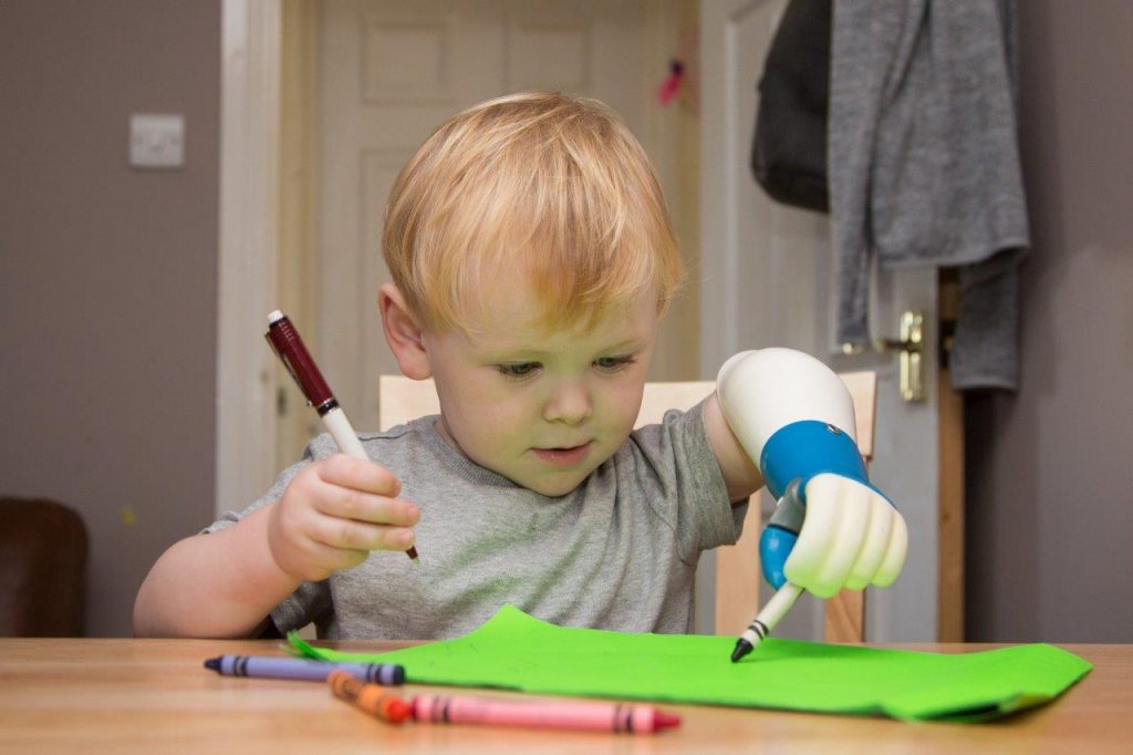
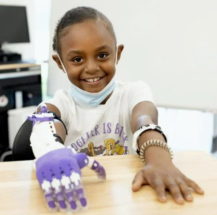
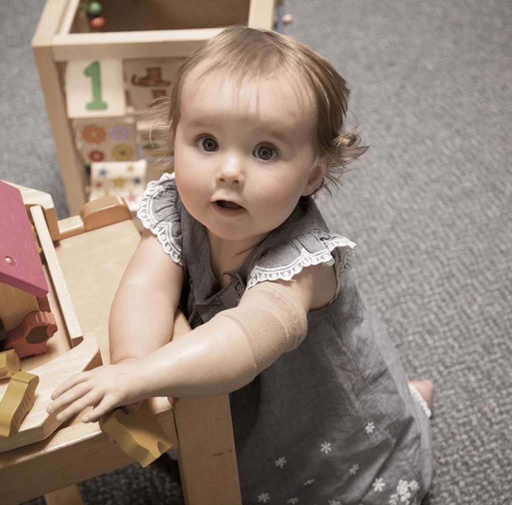
Prosthetic hands are used to support people that are missing their hands due to a congenital condition, an illness or from an injury. They can help with mobility, strength, and everyday tasks. Some children will wear a prosthetic hand throughout their life whereas other children may never wear one. Prosthetic hands encourage children to use both their hands which improves their brain and motor development. They also help with their appearance and self-confidence.

Development of the prosthetic hand

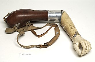
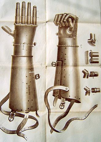
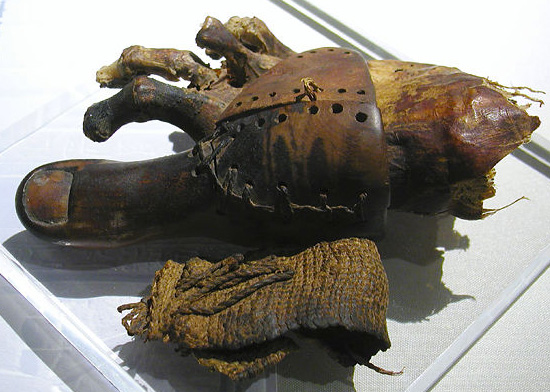
  In the late 15th century, France and Switzerland were making artificial hands. These were made from wood, glue, metal, and leather. In the 16th century, Götz Von Berlichingen wore 2 iron prosthetic hands due to losing his right arm from the war. The second hand was able to hold objects. In the 19th century, William Robert Grossmith created a left prosthetic arm from wood and aluminium.  In the 20th century, plastic was used for prostheses. Today, prosthetic hands are made from silicone, titanium, aluminium, and plastic.

Cosmetic devices

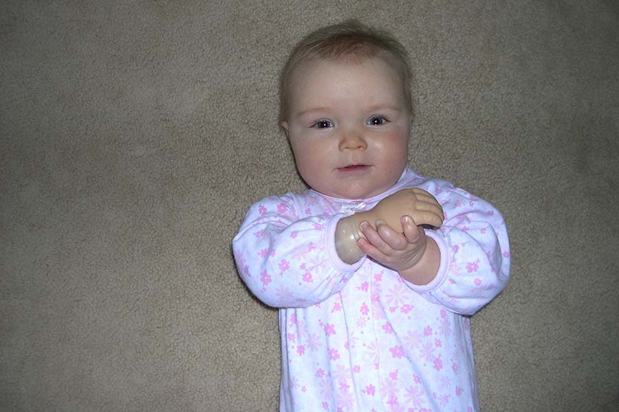
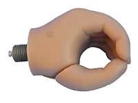
  For children under 18 months an ideal prosthetic hand is a passive prosthetic device which is a cosmetic device. These do not move by themselves; they are made from silicone and are lightweight. The earlier a child starts wearing a hand prosthesis, the more they become accustomed to it. The Greek Series, Infant 2 Hand, L’il E-Z Hand and Lite Touch Biomechanical hands are examples of prosthetic devices that can be used for children.

The Greek Series are suitable from 4 months to 3 years old. They have realistic hand features so they can be used to hold light objects such as small plastic toys.

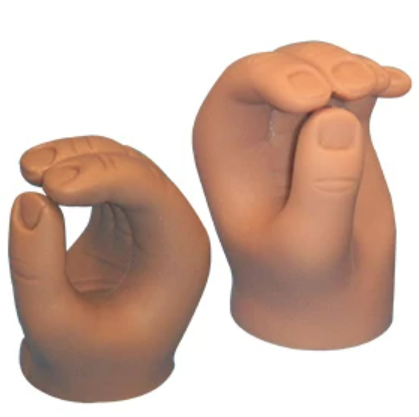
The Infant 2 Hand can be used from 6-18 months. This hand can be used for pushing and pulling objects as the hand is a cup shape.

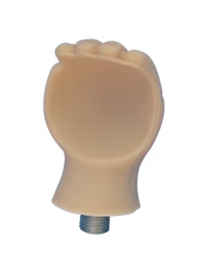
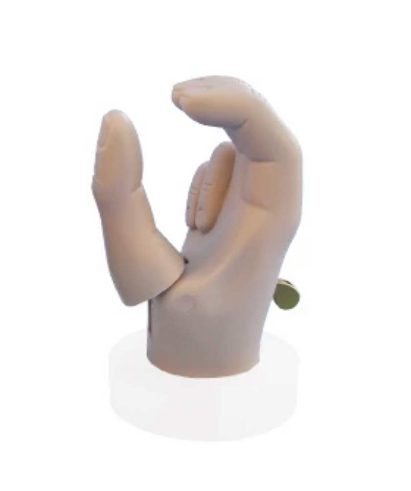
The L’il E-Z Hand is suitable from 6-24 months. This hand has a mechanical thumb which helps infants to grasp objects easily.

The Lite Touch Biomechanical hands are recommended from 2-9 years old. This hand has moulded fingers which can voluntarily open and close.

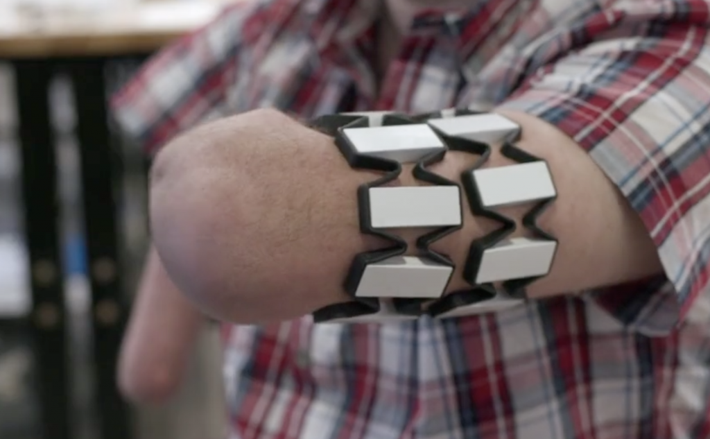
Myoelectric prosthesis for children

  Myoelectric prosthetics are suitable for older children, over 10 years old. These use more advance technology and can benefit children as it develops their muscle memory and helps them perform activities that involve 2 hands.

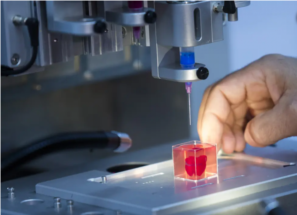
3D printing for prosthesis

  3D-printing technology can make prostheses more affordable for the public. It also reduces the manufacturing time which can take up to 6 weeks. Instead, a prosthetic limb can be created within a day. The aim is to make 3D printing more accessible so people can make their own hands. In 2011, Ivan Owen created the first prosthetic hand using 3D printing. These prostheses are made with plastic, carbon fibre, aluminium or titanium therefore making them lightweight.

Overall, prosthetic hands have developed significantly over the years. They are very important for both children and adults as they can positively impact their lives by developing their everyday skills. There is an amazing foundation called the Douglas Bader Foundation which works with charities in the UK involved in Project Limitless. Project Limitless aims to give all children (that need) a prosthetic arm. Over 300 children have already been provided with one.

Links

https://www.steepergroup.com/prosthetics/upper-limb-prosthetics/hands/trs-paediatric-hands/

https://www.yourkoalaa.com/projectlimitless

AI Bionic Arms Exist: An Innovative Piece of Prosthetic Tech by Atom Limbs


Background on Atom Limbs and Prosthetics

This is the first artificial human arm created by Atom Limbs called the ‘Atom Touch’.

Atom Limbs is a company whose goal is to build a better future for humans by building artificial prosthetics that allow amputees to regain function of lost limbs.

Prosthetics first emerged with the use of materials such as wood and metal, however current technology uses lighter and more durable materials such as carbon fiber and silicon. Despite the modern world having great advancements in technology, most prosthetic limbs have remained the same in that they give the appearance of a functioning limb but are relatively useless to the wearer.

Prosthetic toe from ancient Egypt


Ethical and Societal Considerations

Bionic products tend to be expensive which puts most devices out of reach for many disabled people, who statistics show are more likely to be on the poorer end of society. There may also be supply issues for low income countries who have limited access to specialised medical facilities. This seems to partially be why less than 1 in 5 people with arm loss do not use a prosthetic. So what is the point of making a device to improve quality of life if the intended target audience doesn’t use them?

I read this BBC News article and it appears that Atom Limbs have dwelled upon this ethical consideration positioning their bionic arm around ÂŁ15,000, which – although still a lot – is considerably less than many other similar devices on the market. A more reasonable price for a high quality bionic product. This made me question myself – if I had a missing arm, would I get this bionic arm? Considering its price, my decision would come down to the prosthetics practicality and usability in everyday life. If it functions like a real arm, then it would be justifiable to spend such an amount as it is a life-long investment.


The Underlying Science

The prosthetic arm controlled by your mind | BBC News

Neural Interface

Atom Limbs primarily relies on the use of advanced sensors and machine learning algorithms to interpret signals from the brain and utilise them to move a prosthetic limb. It connects to the wearer’s residual limb with bands of sensors that measure electrical signals produced from muscle movements. Signals are then picked up by electrodes and integrated. This innovative mechanism is known as myoelectric control.

Attachment

The arm attaches via a vest and is a non-invasive procedure which means no surgery is required for it to function. It uses breathable and lightweight materials providing comfort, which I believe would encourage amputees to choose the ‘Atom Touch’ over other prosthetic arms.

Haptic Feedback

One of the challenges posed in the use of bionic arms is the grip strength used on different objects. How would a wearer be able to tell how strongly they are gripping, for example, an egg to ensure it doesn’t crack? The answer is haptic feedback – this is delivered to the user through vibrations which are produced when sensors detect interaction with the environment.

Motion

The near-full range of motion in the elbow, wrist and individual fingers allows the wearer to fully interact with the world and regain their sense of touch. For someone who has not lost a limb, this may seem like a trivial feature, however for an amputee this may be an extraordinary feeling. An amputee reporter from the BBC News operated a digital version of the bionic arm.

“The notion of learning how to control a part of the body I don’t have is almost impossible to describe.”

Paul Carter from BBC News

Printing life : Can 3D-bioprinting organs using stem cells revolutionise medicine?


A 3-D printed heart composed of human tissue from Tel Aviv University.

Medical 3D-printing using stem cells sounds like a dream but recent advances in printing technologies has paved the way for new possibilities in artificial organ printing and regenerative medicine.

Across the globe many individuals who need organ transplants are suffering due to the lack of available donors. The NHS states that in the last 10 years, 1/4 of patients waiting for a lung transplant have died. Utilising 3D-printing to make organs accessible can play a crucial role in removing the issue of waiting years for a donor.

What actually are 3D-bioprinted organs?

They are functional and biologically manufactured replicas of natural body parts, made out of a bio-ink which consists of biopolymers and stem cells which is cultivated in a lab.

I believe 3D printed organs hold promise in terms of transplantation, disease modelling and drug testing, but challenges arise in regards to functionality, availability and if the body will accept the manufactured organs.


How does 3D-bioprinting organs work?

In short, 3D printed organs are made from a mixture of biopolymer hydrogels and cultured stem cells forming a bio-ink. Hydrogels like alginate or gelatin, are used as ‘scaffolds’ in the printing process of the organ. The process sounds simple but researchers must take into account factors like cell/ tissue type and the bio-ink needed for the specific organ.

Mark Skylar-Scott, an assistant professor at the Stanford University department of bioengineering stated the bio-ink is loaded into “syringes and is squeezed out of the nozzle like icing on a cake”. This process is repeated with different cell types, and once complete is provided with oxygen and nutrients. Over time developing to perform its intended function.

Skylar-Scott’s team developed a method to speed up the printing process, it involves printing in clusters called organoids which he describes as a “human stem cell mayonnaise” which is then printed. The video below describes the process in full :

Stanford University/CNET

What role do stem cells have?

Stem cells are old news in the medicinal world, the first successful Hematopoietic stem cell transplant was in 1959! Stem cells have the ability to turn into other cell types and for that reason they are still extremely relevant to this day.

Stem cells are still used to innovate treatments. For example, induced pluripotent stem cells (iPSCs) obtained from de-differentiating skin cells, can turn into any type of cell, making the possibilities for new treatments endless, as stem cells can be acquired without the controversial use of embryos.

The video below shows Dr. Brenda Ogle’s team at the University of Minnesota using iPSCs to create a 3D printed functional human heart pump :

University of Minnesota

The future of 3D-bioprinting organs ?

In 2022/2023, 281 people died while waiting for a kidney transplant in the UK, 3D printed organs can reduce the waiting time for in-demand organs. Wake Forest scientists have been able to grow mini-kidneys and livers, demonstrating the promise that printed organs have for the future .The cost of a liver transplant in the USA is approximately $812,500, 3D-bioprinters that can make printed organs can be as cheap as $45,000, providing a cost effective alternative for patients.

The actual use of these organs is still 20 to 30 years away. Better hardware with the ability to replicate the complex nature of organs, and mitigating issues like biomaterial degradation and tissue integration is crucial if we want to use these organs in the future. I believe that the use of 3D-printed organs can revolutionise medicine and give hope to those in need of transplants.

Cochlear Implants: Tuning and Technology Advancements

Hearing is a sense that I, and likely many others worldwide, take for granted. That said, around 1 in every 6 adults in the UK live with at least partial hearing loss. The technology behind cochlear implants piqued my interest after listening to the insights from users in the lecture, leading me to begin reading parts of Dr Yoder’s cochlear implant journey.

What is a Cochlear Implant?

Cochlear implants are made up of both internal and external parts and require a relatively short surgery to fit. The external parts are made up of a microphone to pick up the sound waves, a language processor to convert the sound waves into electrical signals, and a transmitter to transfer these to the internal components. The language processor and microphone usually rest on the ear, while the transmitter is placed further back on the head. The transmitter is held on via a magnet fitted under the skin during surgery. This allows for the previously converted electrical signals to reach the string of electrodes inserted into the cochlear, which stimulates the auditory nerve directly.

Cochlear implants aid the users’ hearing through a string of electrodes inserted into the cochlea. https://brsh.org/cochlear-implants/

Tuning

One aspect of cochlear implants that I found fascinating was tuning and the period over which it takes place; initial tuning usually takes place one month after the surgery, with periodic sessions after this to slowly ease users into the new sounds. This initially shocked me; “Why does this process take so long?”, I found myself thinking. However, after reading Dr Yoder’s experience with tuning as well as others (seen in the video below), I realised that hearing is not restored with the surgery, but with the practice users do to re-understand how everyday noises and speech sounds. The video below by Cochlear Americas documents four users and their experiences with training themselves to understand how to hear through their new implants, which I found very insightful.

Current and Future Research

A recent publication in Ear and Hearing in 2023 discussed advancements in tuning of cochlear implants, and how artificial intelligence can be used to outperform traditional tuning methods. The traditional method, despite performing less effectively, was preferred by participants, owing to it feeling more comfortable for them. I believe that this aspect of optimising implants should not be neglected; how the users think or feel about such methods, be it tuning or any other aspect of the implants, is of utmost importance.

A less recent article from the engineering department in Cambridge highlights how 3D printing can be utilised to improve cochlear implants. The fluids inside the cochlear ducts are highly conductive to electrical signals and implant users often, due to this conductivity, experience distorted sounds through their implants; this is known as current spread. The use of 3D-printed cochleae, paired with machine learning, allowed researchers to analyse and predict how current spread was impacted by the cochlea’s shape and conductivity. Dr Shery Huang, an associate professor in Bioengineering at Cambridge, suggested that this is an extremely useful application of 3D printing since data regarding patients must be kept private; Dr Huang was quoted in the article, stating that to solve this, “3D printing is a powerful tool to create physical models which might provide a well-characterised training dataset as a purpose-built surrogate to clinical data for machine learning”.

More than just skin deep: advancing the sense of touch in prosthetic hands.

During the lectures on sensors and sensing, we were shown a video of a ‘bionic arm powered by AI’. The video showed a man controlling a prosthetic hand using his mind, and got me thinking about the extent to which a prosthetic hand might be able to replicate the function of a biological hand, in particular if prosthetic hands could ever ‘feel’.

A touchy subject

So much of what we do and how we interact with the world relies on touch. Primarily, touch is important for perceiving pressure allowing you to interact with objects at just the right force. If you pick an egg up with too much force it will break, but not enough force and you’ll drop it. Either way you’ll break a few eggs, but won’t end up with an omelette. There are a range of receptors in the skin which give us our sense of touch. The mechanoreceptors act to convey tactile information from our fingertips to our nerves. Whereas the nociceptors are free nerve endings which conduct stimuli which we perceive as painful. In the past, prosthetic hands could replicate some of the hand functionality, but have not been able to sense tactile information. However, recent technological advances are paving the way for feeling with a prosthetic hand.

E-dermis: the prosthetic skin

The e-dermis designed at John Hopkins.

John Hopkins have developed an engineered material skin, made with fabric and rubber, and implanted with sensors to act as pain and touch receptors. The sensors of the e-dermis can then stimulate the nerves in the residual limb or the amputee through the skin, to allow the perception of both painful and non painful tactile stimuli.

Brandon Preston’s story

Brandon Prestwood implanted with the wires which transmit signals his implanted left arm electrodes The audacious science pushing the boundaries of human touch | National Geographic

After an industrial accident at his work in 2012, Brandon Prestwood lost his lower left arm and hand. After battling with depression after the accident, Prestwood volunteered for experimental research with Cleveland State Western University, leading to the insertion of electrical conductors to the remaining nerves in his residual left upper arm, with four wires then guided up through his residual left arm and out of his shoulder. As the nerves and their link to the brain remain in the residual limb, by incorporating sensors into the prosthetic hand, the signalling can be restored. The sensors in each prosthetic finger convert contact with a surface into an electrical signal, the signal is sent to a computer, then the computer stimulates the correct nerves through the implanted electrodes. By doing this, Prestwood could touch an object with a prosthetic finger and know which finger is touching it.

More than a feeling

You may wonder why the sense of touch is so important. Would it not be easier to shorten the loop, with the sensory receptors of the prosthetic feeding back to an internalised system to modulate the force used by the hand, and forget about transmitting signals to the brain all together? However, the need to perceive touch is for more than just to pick up an egg; touch is also a vital part of being human. From handshakes, to high-fives, to hugs, touching is integral to being human. It’s even engrained in our language, if someone buys you flowers you feel ‘touched’ by the kind gesture. Even Shakespeare alludes to the importance of hands and touch, ‘now join your hands, and with your hands your hearts’. For Brandon Prestwood it was as simple as being able to hold his wife’s hand again with his missing left hand:  “It’s the emotion that goes with any kind of touch. It is 
 it’s being complete.”

Reference links

Prosthesis with neuromorphic multilayered e-dermis perceives touch and pain | Science Robotics

From Research to Reward: Something Lost, Something Gained: High-Tech Prosthetics Build on New Understandings of the Human Body (nationalacademies.org)

New ‘E-Dermis’ Brings Sense of Touch, Pain to Prosthetic Hands – Johns Hopkins Biomedical Engineering (jhu.edu)

The audacious science pushing the boundaries of human touch | National Geographic

Advancements in prosthetics limb technology allow feeling, control | 60 Minutes – CBS News

How Bionic Eyes Have Reshaped the Lives of Visually Impaired – draft

Advancements within bionic devices has boomed within the 21st century, seeing new technologies rescope human augmentation as we know it. The likes of new 3D printers have propelled the boundaries of what we consider possible, in particular enabling the development of new ‘bionic eyes’. The development of this new, groundbreaking technology will likely transform the lives of individuals living with profound blindness; in particular, for those with conditions such as macular degeneration and retinitis pigmentosa which lead to degeneration of retina.

The eye, and the neural signalling which allows us to see is a particularly complex nature. In relatively simple terminology, our eye consists of a retina containing rod and cone photoreceptor cells which help to identify both the intensity and spectrum of light. This can be converted into electrical signalling, and transmitted along the optical nerve into the visual cortex area of the brain to be processed. Various conditions, which lead to the deterioration of this signaling pathway, can be extremely detrimental to individuals sight and their livelihoods.

Thus, the development of this new bionic device is a particularly exciting advancement within the biomedical community. This implementation consists of a electrical macular implant, about the size of 2x4mm and external eye glasses. The electrical component of this device allows stimulation of ganglion cells, whereby electrical currents can be tramistted along the optical nerve and processed in the brain. This technology is currently being developed by a company named SecondSight and is called the Argus II.

One girl who has seen the promising advances in this area is Myah Hauxwell who was born with a rare defect called severe microphthalmia causing blindness . This is a complex condition leading to reduced eye growth, although the precise pathology is still mostly misunderstood. Myah, who is just 9 years old, had undergone 23 surgeries before raising money to have the ocular prosthesis.

To hear or not to hear? That is the question.

A estimated 900,000 people in the UK suffer from server or profound hearing loss (approximately 1.34% of the UK population), and the World Health Organisation estimate that a whopping 5% of the global population suffer from disabling hearing loss! Now you may think that these stats aren’t anything to worry about and that hearing aids will compensate for hearing loss. You wouldn’t be completely incorrect, as hearing aids do help those with partial deafness, but they do not provide the same level of aid for those who suffer from serve/profound hearing loss.

Those who suffer from serve/profound hearing loss are categorised as not being able to hear sounds bellow 71-95dB (for serve hearing loss) and sounds bellow 95dB (for profound hearing loss). To put this into context, imagine you go to a club with a friend who suffers with profound hearing loss. You and your friend would need to stand 1 meter from the speakers in order for them to hear the music, and after a night like that you too may be suffering some hearing loss!

So if hearing aids aren’t the best solution, what is being done? Well in 1961 the first ever cochlear implant (CI) was used, and since then many advancement have been made to shrink the implant and minimise the invasiveness of the procedure. The CI is a device that contains a receiver that is inserted under the skin of the head with an electrode that is implanted into the cochlear directly. Additionally a transmitter and microphone/processor are attached to the receiver on top of the skin via magnets. Essentially what the CI does is receive sound via the microphone and processes that sound into digital information, which passes from the transmitter to the receiver (through the skin) and down the electrode to stimulate the cochlear directly. Allowing for the sounds picked up by the microphone to be heard by the deaf patient.

Diagram showing where the cochlear implant sits within the head.

As of 2022, more than 1 MILLION cochlear implants have been successfully implanted! Granting many people, of all ages, the sense of hearing! So seeing that cochlear implants have the ability to artificially restore one of our key senses, surely no one would object to them? And even if there were, I would assume they’d be ill spirited people that can’t stand the idea of seeing others enjoy hearing. Or perhaps those with personal or religious beliefs that sees the implantation of foreign matter into the body as wrong. Well upon research, there are indeed people as I just described, but the leading voice against CI shocked me and may shock you too.

This voice being those from the deaf community themselves! Many in the deaf community are happy with their lives and don’t see their deafness as a negative attribute. They can communicate via sign language and can still sense the world via touch and sight. So this idea of ‘fixing hearing loss’ can make it sound like those who are deaf are ‘broken’, when they are living healthy lives. In my opinion we, as humans, are designed to be able to hear. So the lack of hearing technically would be a defect, however many of us suffer from different ailments and that isn’t necessarily a bad thing. I understand that for many, deafness is not debilitating, and so do not want to have the CI as they are happy the way they are.

When it comes down to it, cochlear implantations are an elective procedure and if you do not wish to have one you are completely within your rights to do so. However some people still see those who have CIs as “betraying the deaf community” when in reality they are just people making the decision that they believe will most benefit themselves, same as those who opt to not get a CIs.

The Infinite Potential of Tissue Engineering

The potential of tissue engineering.

I find the potential of tissue engineering to be very interesting, mainly because I believe that there is no limit to the potential. One of the main uses for tissue engineering is to replace lost tissue and to maintain damaged tissue or even potentially an entire organ. But that is another issue altogether!

There are some other aspects to tissue engineering, and these include regenerative medicine, which has been used interchangeably with tissue medicine.

The interesting history of tissue engineering

The idea of tissue engineering has always been more of a dream or a fantasy before modern technology came about. A very intriguing perspective that I have found recently explores the idea that the concept of tissue engineering has been around in the early history of man, which is the story of Eve being created from the rib of Adam. This is so fascinating to me because it shows how humanity has this innate desire to create and not be bound by any limitations.

However, in modern times, the concept of tissue engineering was introduced in the late 1980s, and this opened a gate of endless possibilities and different applications for the future.

One of the first examples of tissue engineering being successfully implemented was in 1991, when an individual with Poland’s syndrome was the first human to receive a tissue-engineered implant that was composed of synthetic polymer.

The ability to save human lives.

Unfortunately, in our world currently, there is a shortage of organs that are available for transplantation. Additionally, I was saddened to find an article in the New York Post showing how there are failures in the management of organs and a lot of available organs are actually going to waste and will be discarded even though the number of people that are in desperate need for a new organ will never run out. It was reported that around 17 people die per day while waiting for a transplant, and there are around 106,000 people in America on the waiting list.

This is why I believe that tissue engineering can provide a possible solution to this devastating problem. With the improving technology, the possibility of creating 3D organs is increasing, with biomedical engineering researchers developing 3D temporary organ structures called scaffolds. With this technology, the possibility of creating material that can help with recovery is increasing.

Problems with Tissue Engineering

I am very excited about the prospect of tissue engineering and its potential future applications. I am very aware that there are limitations to this.

Some of these limitations are the materials used. For example, alginate is a difficult material to use as it collapses easily. Moreover, the challenge I believe to be the hardest to overcome is receiving donations from others.

If we were to use stem cells to engineer tissue for another, there are many ethical questions that must be addressed, such as the problems of taking stem cells from a human embryo.

In my humble opinion, I believe that taking stem cells from an embryo is unethical, and this mainly revolves around religious problems, as being raised in a catholic family has shaped my views on certain matters such as this.

However, if improved communication to try and get more people to donate their stem cells, then it may be possible to overcome this problem, and many more lives could be saved in the future.

Here is a video of a scientist explaining his views on the ethical issues of embryonic stem cells that I largely agree with.

Pluripotency in Practice

The maintenance of stem cell lines

Pluripotent cells (PSC’s) are taken from adult tissue or embryonic cells, these can also be induced, known as iPSC’s. This is when the pluripotent stem cell are genetically modified to be reprogrammed. I undertook some work experience at the Sheffield Institute of Translational Neuroscience Lab, which researches Parkinson’s disease, looking at both drug discovery and how the neurodegenerative disease manifests and progresses. Within the research a big task is maintaining different cell lines for future experiments on fibroblasts, induced neural progenitor cells (iNPC’s), astrocytes and neurons; to complete this, cells must be fed with nutritional media to help them grow and split when their is overcrowding in the dish. A stem cell should have the capability of infinite self renewal and differentiation, however in the lab there was a limit for how many times this could be conducted to prevent stress and harm to that cell line. The number of times the cells are split are called ‘passages,’ there was a rough limit of 15 passages per cell line. To ensure continuation of the line for the future, a spare dish is always kept frozen from the splitting process. Some cells used in the lab are brought in already modified for experimentation but other cells needed to be reprogrammed and grown in the lab, this aspect I got to observe during my visit for neuron cells. This entailed harvesting fibroblasts from the skin of a Parkinson patients and control patients with wild type cells which are exposed to a virus. The virus alters the genetic makeup of this cell to allow for differentiation into iNPC’s and eventually into neurons. This process can take anywhere between 2 weeks and 8 months to occur.

The picture on the LHS shows imaging of a stained Fibroblast, this is how the cells will look at start of the reprogramming process. The picture of the RHS shows a view down the microscope of neuronal cells, this is how they should look once reprogramming is complete, new cell lines can be taken and grown from this. Note: if the neuronal cells are taken from a Parkinsons patient they will be induced dopaminergic neuronal cells.

The legality…

There are legal implications with the use of stem cells in the UK, this varies from country to country, whereby scientists need to obtain permission from the governing body, ‘Human Fertilisation and Embryology Authority’ (HFEA) in order to use them. Lawyers, Clinicians, Scientists and Ethicists will determine if the use of that stem cell line is appropriate or not, if granted, regulations require these cells to be stored in the Stem Cell Bank. This bank enables all cells to be overseen, and allows researchers to use existing stem cells if given approval. Here is a link to the UK Government website explaining in detail the protocols put in place in the UK with regards to stem cells.

Ethical dilemmas…

There are many ethical dilemmas surrounding iPSC’s and the regulation of them. iPSC’s are very powerful having the ability to be taken as a fibroblast from the skin and reprogrammed into any cell type including, an egg or a sperm cell, these have been used to create mouse embryos which can develop into fully grown mice. This raises many questions including the implications of this experiment if it was to be conducted on humans in the future, the ownership of the cells – do they belong to the researcher or the donor? Should iPSC’s be used over embryonic stem cells?  Debates are still occuring to determine answers for these questions and come up with a sytem for monitoring embryonic stem cells and induced pluripotent stem cells looking at the benefits and challenges which come with each.

Accreditation:

Hirai, Takamasa, et al. “Country-Specific Regulation and International Standardization of Cell-Based Therapeutic Products Derived from Pluripotent Stem Cells.” Stem Cell Reports, vol. 18, no. 8, 1 Aug. 2023, pp. 1573–1591, www.ncbi.nlm.nih.gov/pmc/articles/PMC10444560/, https://doi.org/10.1016/j.stemcr.2023.05.003.

“Ethics.” The University of Edinburgh, 2 Aug. 2021, www.ed.ac.uk/regenerative-medicine/about/ethics.

“Sheffield Institute for Translational Neuroscience.” Www.sheffield.ac.uk, 10 May 2023, www.sheffield.ac.uk/sitran. Accessed 8 Mar. 2024.

Will Amniotic fluid stem cells be the future of organoid generation and congenital disease prevention?

Congenital diseases are estimated to be the cause of death for 240,000 newborns within the first 28 days of life and a further 170,000 deaths of children from the ages of 1 month to 5 years old according to statistics generated by the WHO. Identification of these diseases at an early stage Is therefore vital in giving the best chance to apply the most effective treatment whether that be surgery or pharmaceutical therapeutics.

Scientists have had a difficult time isolating tissue-specific stem cells derived from the foetus due to their limited sample obtainability. Collection of foetal tissue has to be done in regulation with ethico-legal restrictions laid out by the Human tissue act of 2004. The extraction of foetal tissue cannot be done past 22 weeks of pregnancy and is usually carried out a post-mortem. The isolation of foetal stem cells has sparked large ethical debates due to the harm caused to the foetus which usually results in the termination of pregnancy. Despite this, these stem cells are crucial in understanding the late-stage development of the foetus and can aid in discovering the severity of cognitive diseases which has a great potential to save many lives.

However the ethical debate may come to an end, a recent study published by ‘Nature Medicine details a groundbreaking revelation into a new method of foetal stem cell isolation through the use of amniotic fluid, the yellow-tinged liquid surrounding a baby during development with the main role of protection. It was found that these foetal stem cells leak into the amniotic fluid during nutrient and urine turnover. This would mean that the direct extraction of stem cells from the foetus will be eluded. The scientists at the Great Ormond Street Hospital and University College London could isolate progenitor cells within the fluid by a technique called fluorescent activated cell sorting or ‘FACS’ along with a range of other techniques to fine-tune the culturing conditions. The cells grew many variations of organoids such as small intestines, lungs, and kidneys in both the progenitor form and epithelia variations. The organoid development will coincide with the development of the organs in the late stage of pregnancy and hence enable us to study the organs in vitro whilst allowing the foetus to continue development. This in-vitro work will enable real-time organoid models representing the current stage of organ development allowing us to recognise and analyse the signs of congenital diseases such as CDH (the condition in which there is a hole formed within the lungs) and hence allow us to come up with personalised pharmaceutical or surgical treatments to counteract the defect by targeting the transcription and genomic expression of the individuals organoid.

This new research has allowed us to understand late-stage development during pregnancy for the first time past the 22-week threshold. The study showed that those who are developing the CDH condition showed a difference in gene and protein expression and hence altered development compared to the healthy organoids. The production of organoids through the foetal stem cells has allowed treatment reflection determining the effectiveness and efficacy of the current treatments available and has allowed the possibility to test new treatments in a less restricted manner.

Currently, the research carried out has led to developments in CDH, TTTS, and MMC treatment however the research showed that within some conditions, improvement has been limited. LUTO stands for ‘lower urinary tract obstruction’. One way this can be treated is by providing a ready supply of amniotic fluid hence removal of the amniotic fluid for generating organoids for further study may be risky hence fewer samples have been obtainable and so the cause of LUTO is still unknown with lack of definite treatments on the horizon due to a variety of isolated cases of the disease.

Overall, I believe this new research has ultimately paved the way to view the late-stage development processes to a further extent than ever before and is an innovative approach that could lead to the almost complete eradication of a whole range of congenital diseases which has the potential to benefit whole spectra of individuals and their families whilst also bypassing one of the most major ethical science debates.

The link to the original nature science paper is here!