The University of Southampton

Engineering Replacement body parts 2023-2024

An interdisciplinary module

Comparing Behind-the-ear and Off-the-ear Cochlear Implants of UK Brands

Cochlear implants have been an approved method of treatment for the profoundly, and more recently severely, deaf since the late 20th century. Their continued technological improvements since have provided those patients with the ability to hear through a processor (Hainarosie, 2014). The audio is not a perfect replication of natural hearing, but allows for interpretation of speech and sound in a way the brain can understand. Modern technology around cochlear implants provides patients with the option to have the external processor of their implant either behind their ear or off of their ear. Both designs allow for bluetooth connection between phone and processor, and each option has its own positives and negatives for users to consider.

Behind-the-ear (BTE) Cochlear Implants

BTE cochlear implants are the originals – this design for the external processor has been used since implantation in the cochlear began. The processor (containing the microphone) sits on the ear of the user and a short cable connects this to the magnet that transfers the audio information to the electrodes implanted in the cochlear. The location of the microphone on these processors differs between companies. For example, Advanced Bionics’ Naida Cl M sound processor has the microphone dipping down into the outer ear region whereas Cochlear’s Nucleus 8 sound processor has dual microphones. Each company has taken a slightly different approach to the goal of reducing background noise and making the sound as close to natural hearing as possible.

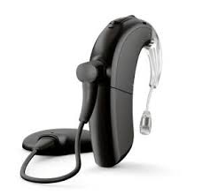
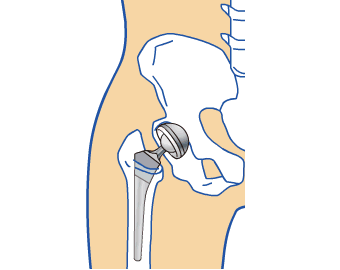
BTE – Naida Cl M sound processor – Advanced Bionics

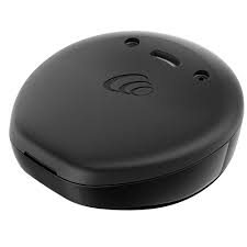
OTE – Kanso 2 sound processor – Cochlear

Off-the-ear (OTE) Cochlear Implants

OTE cochlear implants are a more recent development in cochlear implant technology with Cochlear’s Kanso 1 sound processor being released in 2016. These processors contain only one piece that sits on the side of the head connecting directly to the magnet inside the head. There is no part that rests on the ear. Because of this, some users find it more comfortable because their ear can have a break from holding the processor. However, it is often found that the magnet needs to be stronger to ensure that the processor does not fall off because it is a less secure connection. This is a problem for some people as they feel more comfortable with a processor on their ear when they are playing sport or in other situations where the processor could get dislodged. The single piece design of the OTE processor means that the microphone is placed on the side of the head. This can have some impact on the audio that the user receives as it is not being collected from the natural location – the outer ear.

Conclusion

Some cochlear implant users opt to obtain both a BTE and OTE sound processor after surgery so that they can use each to their strengths. Throughout reading for this blog I found that my preferred design is the Naida Cl M sound processor by Advanced Bionics because of its microphone location and BTE design. A BTE design allows for better microphone placement and a more secure feeling whilst an OTE design allows for a more discrete processor with fewer pieces attached to the head – perhaps making glasses or hat wearing slightly easier. To improve comfort around OTE sound processors, there are clips and headbands available. I think that it is important for each user to be able to weigh up the pros and cons of each processor type to make an informed decision about which would work best with their lifestyle. Perhaps the difference in age between two patients would be enough to result in different choices. Access to both processor types appears to be a great solution for those who find that OTE and BTE processors are each useful in different parts of their life.

Further reading suggestions:

A Reddit thread discussing personal experiences with OTE and BTE processors:

https://www.reddit.com/r/Cochlearimplants/comments/wtdkvd/ote_kanso_2_vs_traditional_bte_processors/?rdt=42393

A list of current cochlear implant processors available in the UK:

https://www.bcig.org.uk/ci_manufacturers.aspx

Cochlear’s comparison of their current sound processors:

https://www.cochlear.com/us/en/home/products-and-accessories/cochlear-nucleus-system/nucleus-sound-processors/compare-nucleus-sound-processors

References:

Hainarosie, M., Zainea , V. and Hainarosie , R. (2014) ‘The evolution of cochlear implant technology and its clinical relevance’. Journal of Medicine and Life. Available at: https://www.ncbi.nlm.nih.gov/pmc/articles/PMC4391344/ (Accessed: 8 March 2024).

Waiting for a knee replacement

Photo of Doctor holding a post-op knee

My dad, along with almost 400,000 others, has been waiting for a knee replacement on the NHS for over a year [1]. This wait has caused numerous complications for him, as originally he was only going to need a partial knee replacement. Now a full knee replacement is required, which will drastically increase recovery time, pain levels, complications and the cost of the procedure on the NHS [2].

Partial knee replacements have been shown to improve quality of life and could of saved almost £2000 on the NHS over my dad’s lifetime if they acted sooner [2]. If we were to go private, a full knee replacement would cost around £14,000 [1]. This amount of money is unfeasible for a lot of UK citizens, especially since the cost-of-living crisis. But for those who cant pay up, they have to suffer with degradation of health and lifestyle.

Image showing partial knee (left) and total knee (right) replacement https://www.drsantoshshetty.com/partial-vs-total-knee-replacement/ (accessed 11/03/2024)

Knee replacement surgery is rapidly developing, but is only accessible to those paying privately. By choosing NHS, you will lose access to using CT Scans to create 3D images of the patients knee, having assisting robotics in surgery and less invasive procedures. All of these have the potential to decrease recovery time and increase surgery success [1]. I believe the NHS should offer a partial payment service for those undergoing knee replacement, as some individuals who cannot afford ÂŁ14,000 for private may still like to pay extra for more modern materials or procedures in order to increase their quality of life. This offers more balanced healthcare across those with different incomes and allows all individuals to have a say on how they want their body to be treated.

Day-to-day life becomes much more difficult; walking is a challenge, so chores around the house become an impossible task. Some individuals may even need to take time off work or find a new job entirely. Exercising can also become difficult, which may lead to weight gain. However increased weight leads to more pressure on the knee, creating higher levels of pain and will make recovery even harder after surgery.

Knee issues can be incredibly isolating and have a massive impact on mental health. It’s important to note how much waiting lists impact mental health. My dad has stated that the stress and pain that he’s gotten from his knee has worsened his heart condition – yet another issue that has arisen from having to wait. He’s also unable to play with his grandsons, as even getting onto the floor is an impossible task.

Treating patients for knee pain also becomes difficult with long waiting lists. Knee pain is excruciating, but there are no pain killers that are designed to be taken for moderate-severe pain daily for over a year.  This limits any help to only walking stick and physio exercises in the hopes of reducing pain.

Current NICE Guidelines state that adults should be allowed a choice between partial and full if both options are suitable [3]. But with increasing waiting lists, the “choice” is made redundant. With no end in sight, my dad’s knee will continue to degrade, leaving him with increasing pain with each passing day.

Recommended watch – Dame Judi Dench’s story on her total knee replacement

References:

1.           Knee Replacement Surgery in 2023: Should you Stick with the NHS or go Private? 2024  11/03/2024]; Available from: https://www.thebestofhealth.co.uk/health-conditions/consultants-specialists/how-much-does-knee-replacement-surgery-cost-in-the-uk/.

2.           Burn, E., et al., Cost-effectiveness of unicompartmental compared with total knee replacement: a population-based study using data from the National Joint Registry for England and Wales. BMJ Open, 2018. 8(4): p. e020977.

3.           Joint replacement (primary): hip, knee and shoulder. 2022, NICE Quality Standard 206.

Stem Cells, Ethical or just Practical?

I recently reviewed an interesting article highlighting the ethical implications associated with Stem cell research. It highlighted a common pitfall – highlighting the promise of Stem Cells to outweigh the ethical factors associated with them.

Generally when I think about medical advancements and promise, I tend to go straight to stem cells of all natures, ranging from unipotent, mulitipotent and pluripotent stem cells. They all have adept functions and are involved in numerous clinical trials throughout many different cohorts of medical professionals. They are involved in treating a range of disorders ranging from neurodegenerative disease to traumatic injuries which have lead to the deterioration of muscle tissue… Do you see how easy it is for me to talk about the science behind Stem Cells rather than the ethical implications? This is a commonality I have seen throughout my wider research when trying to learn more about Stem Cell ethics.

I know what you’re thinking, excluding the ethicality, this all sounds very promising… and truly it is, however, do you think Stem Cells are being used appropriately? Whatever the answer to that question is, let me lend you some information to reflect on.

The Peer reviewed clinical trials being done on diseases such as Parkinson’s and Alzheimer’s disease are incredibly promising and have shown amazing results… integrated amongst highly scientific jargon. What is not amongst this jargon is what the other uses of Stem Cell include and how much the treatment costs.

Stem Cell research is novel and people suffering from these disorders can undergo clinical trials for the price of normal healthcare, however, this requires a complex selection process so that scientists can pick specific patients in which they believe their treatment might be most effaceable rather than for those who need it most. Additionally, Stem Cell treatment outside of clinical trials is offered to the highest bidder. Personally, I believe Stem Cells are going to be amazing in the future, however, in the current climate, I cannot condone them for a number of reasons! But to name a few:

These treatments require much more testing and evidence to support their beneficial effects as oppose to their negative effects and those who buy into relatively untested Stem Cell treatment programmes might do themselves more harm than good whilst funding the growth of untested treatments. Those who need it most do not have it readily available to them, rather, it goes to the highest bidder. Therefore, a last ditch attempt to save a patients life may be overlooked by scientists wanting more positive outcomes or as they have been shown a bigger pay cheque by a patient and/or their family!

It also supports the genetic editing and creation of babies of a high compatibility to children with a disorder requiring Stem Cell transplants. These are known as saviour siblings who may be born into a life or surgery and hospital treatment. How is that fair? They don’t deserve to be thrown into a life of suffering and torture! This list is endless, however, finally I will touch on the doors that are opened through this Stem Cell research. Genetic engineering is closely linked to Stem Cell research and further advancements in one field will promote advancements in the other. How far out are we from couples with the most money being able to develop the “perfect child”?

Is a medical treatment which goes to the highest bidder and can jeopardise the lives of the youth of tomorrow ethical? Or is it just lucrative?

A regenerative approach to reverse menopause?

As an aspiring Clinical Embryologist, I was particularly interested in our lecture on tissue engineering and promoting tissue regeneration. In the reproductive system, ovaries are generally the first to suffer decline in function over time. With a growing number of modern women seeking to beat age-related declines in fertility levels, there is much potential in using regenerative medicine strategies to restore ovarian function and overcome infertility. In 2016, success in a new experimental technique to increase fertility potential was published, using platelet-rich plasma (PRP), termed ovarian rejuvenation.

How does ovarian rejuvenation work?

PRP treatment has been used to treat osteoarthritis and joint degeneration. It involves separation of a patient’s blood sample via a centrifugation process to remove red and white blood cells to obtain a concentrated sample of plasma with a 5 to 10 fold higher concentration of platelets. Platelets are well known for their blood clotting abilities; however, they also facilitate tissue regeneration. Platelets contain granules which deliver various growth factors upon activation, including vascular endothelial growth factor (VEGF), platelet-derived growth factor (PDGF) and fibroblast growth factor (FGF). These promote vessel wall permeability and proliferation of endothelial cells and fibroblasts. Since 2016, interest has grown for applications of PRP in reproductive biology, where direct injection of PRP into the ovary has been reported to induce hormone stabilisation and resumption of menstrual cycles in infertile menopausal women.

Figure 1. Process of ovarian rejuvenation (https://www.ivf-spain.com/en/regenerative-fertility-medicine/)

UK regulations

Whilst ovarian PRP treatment is actively in practice in countries such as Greece, USA and Turkey, it is not currently approved for in the UK by the Human Fertilisation and Embryology Authority (HFEA). Additional therapies and techniques such as ovarian rejuvenation are classified by the HFEA as fertility treatment ‘add-ons’. Although add-ons aim to improve fertility treatment outcomes, the evidence to support their efficacy and outcomes is usually missing, limited or lack reliability. With the UK fertility sector being a competitive market and where 60% of treatments are privately funded by patients, the HFEA is concerned that add-ons are offered for commercial interests rather than as best practice for patients. In December 2023, the Progress Educational Trust held a conference on ‘Updating Fertility, Embryo and Surrogacy Law’ where the HFEA proposed implicating fines as sanctions to fertility clinics for mis-selling of add-ons. Case studies of ovarian rejuvenation appear to present encouraging outcomes, however, there are often limitations in their experimental design such as by absence of sham injection groups or randomisation. The duration of effect after ovarian PRP is unknown, and potential short- and long-term risks are not yet understood. A large-scale, controlled randomised clinical trial is required to confirm efficacy of the therapy before ovarian rejuvenation may be introduced into UK clinics.

Ethical considerations

Ovarian rejuvenation has potential to improve chances of older and menopausal women conceiving their own biological child. However, some debate whether it is ethically acceptable to become a mother at an advanced age, given the likelihood of medical and health-related risks for both the mother and offspring.

I believe that such views are culturally skewed by ageism and ableism, leading to portrayal of older mothers as deliberately risk-taking when infertility and age of motherhood arises from life circumstances outside of a woman’s control. I hope that sufficient research may soon be conducted to prove efficacy of ovarian rejuvenation and its related regenerative therapies in fertility.

References

Pantos K. et al. Ovarian rejuvenation and folliculogenesis reactivation in peri-menopausal women after autologous platelet-rich plasma treatment: https://sa1s3.patientpop.com/assets/docs/111052.pdf

HFEA, The responsible use of treatment add-ons in fertility services: a consensus statement: https://www.hfea.gov.uk/media/kublgcp3/2023-10-19-treatment-add-ons-consensus-statement.pdf

Louisa Gheveart Associates, Fertility Law Reform In The UK: How Much Change Do We Want?: https://louisaghevaertassociates.co.uk/fertility-law-reform-in-the-uk-how-much-change-change-do-we-want/

Metal implants: Good or Bad?

A metal implant is a biomaterial commonly used in orthopaedic surgery to help bones heal, or replace them entirely. Alloys are most typically used. They are designed to be non-corrosive, hard and durable – everything needed from the implant. In the photo below, the hip replacement one is probably the most recognisable, which 71,000 people in the UK have.

https://www.sciencedirect.com/science/article/abs/pii/S1286011518302479

Deciding on the make of the implant is very important. An article I read discussed how doctors need to consider what metals the patient has already been exposed to and what metals will be problematic, however a rejection can occur without any previous hypersensitivity. It suggests management strategies in the case the implant causes immune rejection. It states “Successful medical management with oral atropine sulphate has been reported in a patient with titanium pacemaker as well as with oral corticosteroids in a patient with titanium bioprosthesis for a spinal fracture”. This highlighted the importance of figuring out what metal to use in an implant, something I had not considered in much detail. The following link describes some types of implants: https://youtu.be/FfRZuNaKGdU?si=dT2X0tKhaC6ayGcF.

Image showing examples of types of implants

Patient’s reaction to the metal implantation varies dramatically. Some people show no rejection of the metal, others can show hypersensitivity to the metal and their body actually rejects it, causing intense pain and inflammation for the individual, potentially to the point that the implant has to be removed, but then what do they do to fix the issue?

Fixing the issue, but not completely…

Something which is used to reduce the risk of implants failing is covalently immobilising biomolecules onto the metal surface. An article in the Biomedical Engineering Advances journal believes that using a covalently attached immobilised biomolecule or not is a main determinant in the efficiency of the implant. Of course, this depends on what biomolecules are used and the patient’s individual differences, everyone’s immune system will react differently to the same thing. An example of an immobilised biomolecule is fibronectin, this forms an amide bond with the metal surface, others investigate Arg-Gly-Asp-containing peptides and ubiquitin.

A downside to the use of metal implants is stress shielding. This occurs when the bone density decreases as the stress load on the bone is reduced, due to the presence of the implant. This is a major issue for implants that will not be lifelong (a lot of implants – they do not last forever). This issue may not be apparent until after the metal implant is removed, such as after a severe femur break and the leg is significantly weaker and smaller.

So, what happens when metal implants do fail?

Well, in the UK, patients who’s implants fail, or cause harm, are able to bring legal action against the manufacturers and they are held accountable. The Nuffield Council on Bioethics in June of 2019 estimates ‘over 300 UK patients whose hip implants had failed brought legal action against the manufacturer under the Consumer Protection Act 1987’.

My opinion?

I believe there are lots of ethical considerations that need to be discussed at greater length. Since metal implants have lots of uncertainty surrounding their lifetimes and consequences (as most of it has not yet been seen) it makes it difficult for patients and doctors to make the correct decision, however this does not mean informed consent is dismissed.

The Complications Caused by Implant failure

My Experience with Implants

What’s every teenager’s worst nightmare? To me, it was being told at the young age of 13 that I was missing 2 adult teeth! But why was this such a bad thing? Because the corresponding milk teeth would eventually become too weak, fall out, and nothing would grow back. 9 years later brings us to now, and unfortunately, that dreaded day came. Luckily for me, the medical world has advanced, and dental implants exist, meaning I don’t have to be ‘toothless forever’, or so I thought. 

Recently, 1 out of my 2 implant screws decided it was going to fail and fall out causing a delay in receiving my new teeth. I felt this massively inconvenienced me as I now must remain ‘toothless’ for a little longer. However, this experience had me thinking about the bigger picture of implant failure and I realised that my implants were only for cosmetic purposes. What if my implant was for my knee or hip and that was to fail? How much of an inconvenience would that be? Intrigued, I decided to dive deeper into the topic.

Implant Failure in Joint Replacements

From reading this article by Steven Richard Knight et al, I’ve learnt that, amazingly, hip replacements date back to over 100 years ago when the first surgery took place in 1891 in Germany. Ever since then total joint replacement has advanced in the materials being used, the surgical techniques and technology. This has helped with the life expectancy of the implants which ultimately has reduced the percentage of failure seen today. Although it’s not foolproof yet. 

Joint replacements are mostly seen in people over 60 due to factors such as osteoarthritis. However, they aren’t uncommon in the younger generation.  In a study by Lee E Bayliss et al, I found it interesting that you are more likely to experience implant failure if you’re younger. After further reading, I started to understand why this is the case. Imagine living a day in the life of a 20-year-old and then a day as an 80-year-old. I’m sure you will agree that these days are substantially different. At 20, you’re going to be more active which causes the implant to be put under more strain. This causes the bone to wear down causing it to loosen and fail. However, aseptic loosening isn’t the only reason for implant failure. Other factors such as infection can also be a cause which you can find out more about here.

The Complications of Major Implant Failure and How it is Resolved

When my implant was failing it was obvious to detect as it started to rise out of my gum until it eventually fell out. However, I began to wonder how you would be able to tell if an implant placed inside your bone was failing.  I did some research on the symptoms and it explained how you would experience severe pain and instability in that joint. Imagining what that would be like I decided that although I felt very inconvenienced, it was minor compared to this. These patients would have to endure 1-2 more surgeries to receive joint revision and I only have to wait for the bone to heal before replacing my dental screw. However, even though implants come with a risk of failure, I think it’s incredible that doctors can fix these problems and I think it’s a risk worth taking.  

Ocular Prosthetics – What Can We See In The Future?

Our eyes are one of the most complex sensory organs in our bodies and are taken advantage of everyday. The geometry and intricacy of our eyes is so specific, sometimes you wonder how biology came up with it! In cases of impairment or complete absence of an eye/eyes, we need to find ways to bring back function effectively as well as making them as realistic as possible, to allow for better quality of life. Prosthetic eyes have been around for millennia, but mainly for the purpose of restoring aesthetics. It’s time to further develop the functional aspect.

Currently…

Traditional ocular prosthetics were typically made of acrylic but now use silicone, or a combination to mimic appearance and natural eye movement. By attaching the prosthetic to residual eye muscles, its allows for limited functionality. Aesthetics have come a lot further in terms of mimicking the natural eye by customising things like colour of the iris, iris patterns and blood vessels. This type of customisation may make patients feel more confident in their prosthetic and further enhance their quality of life. As far making and implanting these prosthetics, the procedure has become easier and more sustainable by using 3D printing and quicker, less invasive surgeries. Research has been conducted to incorporate electronics to give the user rudimentary vision, using things like built-in cameras with sensors, which detect and light and dark. But how can we develop this further?

In The Future…

Loss of vision can come in various forms, e.g it maybe congenital, damage caused by injury or even cancer. Therefore, we must consider the best options which take into account risk of surgery, what patients expect from their implant and if it is the most suitable option. With all these things in mind, advancements in ophthalmology and neural circuits can eventually lead to enhanced functional capabilities of ocular implants. What areas of research does this include?

Curing Blindness using a Bionic Eye, Future Now
  • Neural Interface : Involve incorporating cameras, sensors, and neural interfaces to transmit visual information directly to the visual cortex of the brain, by bypassing components that would otherwise be needed to transmit visual information, for example damage to the optic nerve. This is the type of technology we see in the development of a bionic eye.
  • Biocompatibility: Improvements in materials science could lead to the development of ocular prosthetics that are even more biocompatible and long-lasting, reducing the risk of complications such as infections, tissue rejection and the risk from additional surgery.
  • Regenerative Medicine: Bioengineer artificial fully functioning eyes that are indistinguishable from natural eyes. Using stem cells and other biological materials to create new tissue or make completely artificial eyes, engineered using nanowire to replicate photoreceptors and silicone to replicate the vitreous humor in our eyes. This type of artificial eye has been developed and may surpass the ability of an actual human eye.
  • Artificial Intelligence: Integration of artificial intelligence could enhance the functionality of ocular prosthetics by providing features such as automated image processing, object recognition, and augmented reality overlays, further improving the wearer’s visual experience.

Summary

As scientists, there is much we must take into consideration. Managing patient expectations, improving quality of life but simultaneously making it accessible to everyone. Advancements like these can be heavily capitalized to make a profit, which then creates inequality and reduces accessibility to those who need it most so innovation must come with caution. The future of ocular prosthetics holds promise for significant advancements in both aesthetics and functionality however, it’s important to note that many of these developments are still premature and may take time to implement.

Pre-natal “organoids” – the future of treating congenital disorders?

Congenital disorders (more commonly known as congenital birth defects) contribute to a large portion of paediatric disabilities, and can persist into adult life, if the patient survives that long. The World Health Organisation (WHO) estimates that 240,000 newborns per year die around the world within 28 days of birth due to congenital birth defects. If the affected child survives beyond this stage, a further 170,000 children aged between 1 month and 5 years will die as a result of congenital abnormalities. So, why are these numbers so high? And what can be done to reduce them?

Video from the Centers for Disease Control and Prevention (CDC) explaining congenital birth defects.

As congenital disorders are usually identified after birth, when the abnormality has truly developed and “set in”, treatment aims to improve quality of life rather than cure the patient. Until now!

A “mini kidney” produced from stem cells in the amniotic fluid. Immunofluorescent staining reveals the presence of kidney-specific markers (e.g. GATA3 (distal tubule marker), LTL (proximal tubule marker) and ECAD (apical cilia marker)).

Scientific researchers from University College London (UCL) and Great Ormond Street Hospital (GOSH) have successfully extracted embryo-derived stem cells that are circulating in the amniotic fluid of late-stage pregnancies (i.e. up to 34 weeks), and developed miniature organs (so-called “organoids”) from these cells. Previously, foetal sampling in the UK has only been permitted up to 22 weeks after conception (which is the legal deadline for termination), hindering the ability to study late-stage foetal development. As the stem cells collected by the researchers are sourced from the amniotic fluid, rather than the foetus itself, it allows a ‘bypass’ of the legislation.

The research paper, published this week (March 4th 2024) in Nature Medicine, explains in detail the process of collecting stem cells from the amniotic fluid non-invasively, and culturing epithelial organoids that exhibit features reflecting their tissue of origin (i.e. small intestine, kidney and lung).

Video by Rajamanickam Antonimuthu explaining the process and potential of stem cell-derived ‘organoids’ in prenatal medicine.

This development is extremely exciting for prenatal medicine! The work provides a new opportunity to study late-stage foetal development (something which has not been possible before now), furthering knowledge and understanding. The organoids can also be used to model congenital disorders, extending knowledge on these, particularly in the later stages of development. Researchers now have the potential to develop new methods of diagnosis, prognosis and personalised therapy for congenital disorders, all because of a tiny little organ!

I find this research inspiring and highly interesting, as well as hugely promising for prenatal medicine. Anything that can help reduce the morbidity and mortality of congenital disorders is a huge step in the right direction. The researchers have since expressed their vision to extend the method into the production of organoids from mesenchymal and haematopoietic tissues, allowing the treatment of a wider variety of diseases. I am excited to see how this develops, and, who knows, maybe these tiny organs will be the key to curing congenital birth defects!

Stemming the tide : How stem cells could kick MS to the curb – draft

Stem cells represent a groundbreaking frontier in medicine, offering a revolutionary approach to treating a myriad of diseases and injuries. At the core of their promise lies the remarkable plasticity and self-renewal capacity of stem cells. Unlike specialised cells in the body, which have limited regenerative capabilities, stem cells retain the ability to proliferate and differentiate into specialised cell types, such as neurons, muscle cells, or blood cells. This remarkable versatility makes them invaluable tools for repairing damaged tissues, replacing dysfunctional cells, and restoring organ function in a wide range of conditions.

One of the most compelling applications of stem cells lies in regenerative medicine, where they offer hope for individuals suffering from degenerative diseases and injuries such as MS. Multiple sclerosis is a chronic autoimmune disease of the central nervous system (CNS) characterised by inflammation, demyelination, and damage to nerve fibers. It is believed to result from a combination of genetic predisposition and environmental factors triggering an abnormal immune response. MS typically presents with a variety of symptoms, including fatigue, weakness, numbness, vision problems, and difficulties with coordination and balance. The course of the disease varies widely among individuals, with periods of relapse (exacerbations) followed by partial or complete recovery, and periods of remission. Over time, however, MS can lead to cumulative neurological damage, resulting in permanent disability. Treatment aims to manage symptoms, reduce the frequency and severity of relapses, and slow disease progression through medications, rehabilitation therapies, and lifestyle modifications.

Over 2 million people worldwide suffer from MS and before now treatment has focused mainly on symptom management and not the initial problem. A person develops MS when their body’s own immune system attacks the myelin, an insulating and protective sheath, that surrounds nerve fibres. This causes the disruption of messages sent around the central nervous system. The central nervous system consists of the brain and spinal cord. The particular section of the immune system that initiates the attack are cells called macrophages that eat harmful cells or pathogens that have made their way into the body. The macrophages found in the brain are called microglial cells and in progressive forms of MS they cause chronic inflammation and damage to nerve cells.

Now, in research published in the Cell Stem Cell, scientists have completed a first-in-human, early-stage clinical trial that involved injecting neural stem cells directly into the brains of 15 patients with secondary MS recruited from two hospitals in Italy. The trial was conducted by teams at the University of Cambridge, Milan Bicocca and the Hospitals Casa Sollievo della Sofferenza and S. Maria Terni  (IT) and Ente Ospedaliero Cantonale (Lugano, Switzerland) and the University of Colorado (USA).

The stem cells utilized in the study were derived from brain tissue sourced from a single miscarried foetus, these stem cells would be classed as adult stem cells. This method offers a solution to the practical hurdles associated with sourcing foetal tissue from multiple donors. Over a span of 12 months, the Italian research team closely monitored the patients and observed no treatment-related fatalities or severe adverse effects. While temporary or reversible side effects were noted, none of the patients experienced a deterioration in disability or symptoms throughout the study duration. Moreover, there were no indications of relapse symptoms reported, and cognitive function remained relatively stable. As a result, the researchers concluded that the patients demonstrated considerable stability in their disease progression, showing no signs of deterioration. However, the initial high levels of disability among participants present challenges in conclusively affirming the findings.

There are some ethical implications of using stem cells from a miscarried foetus (need to do more research on harvesting stem cells in this instance and not just from embryos)

Need to reduce word count / sum up sections more

How 3D printed prosthetic limbs became the newest revolution in medicine

A 3D printed prosthetic arm example design

Prosthetics are substitutes for missing limbs in the body, in particular arms and legs, but also bones, heart and arteries. There are 30 million people in need of replacement limbs, but the main challenge that people face is that prosthetics are very expensive.

According to this report, the average cost for a single prosthetic limb is around $4,500 USD and can go as high as $50,000 USD. For people in low income countries, this can be unattainable and for people in high income countries, this can be expensive. As a result, I started to think about what solutions are out there that could potentially help this cause. After some research, I came across the field of 3D printed prosthetics, which caught my eye.

What exactly are 3D printed prosthetics?

Essentially, 3D printed prosthetics involves using additive manufacturing methods such as 3D printing to create artificial limbs instead of manually manufacturing them. 3D printed prosthetics are composed mainly of plastic, just like traditional prosthetics, but can also use materials such as acrylonitrile butadiene styrene, or simply ABS, plastics, as well as bridge nylon for a stronger material.

There are 4 main types of prosthetics: transradial, transhumeral, transtibial, and transfemoral. The first 2 types are implants above and below the arm, whilst the last 2 types are implants above and below the knee. Additive manufacturing enables the fast production of these implants.

How do 3D printed prosthetics work?

3D printed prosthetics offer a more streamlined approach to the manufacturing process of prosthetics in comparison to the traditional method. There are 4 steps to this approach, with the first step being 3D scanning. This involves using medical imaging methods such as X-rays and Computerised Tomography (CT) scans to collect images of the patients broken limb.

The next step involves the images being modelled by prosthetists to create and design the required device. This is heavily influenced by the level of detail the computer software provide. The third step is the 3D printing itself, which print layers of the material to create a bonded object. This method prints lots of complex structures in a short period of time. Finally, the device made is fitted onto the patient, where the device made is designed to match the patients anatomy.

This video details the company Unlimited Tomorrow, who create prosthetic arms using 3D printing which has led to the production of their True Limb device.

Freethink/YouTube

Benefits and Challenges of 3D printed prosthetics

The benefits that 3D printed prosthetics provide are the reductions in the manufacturing time and costs of the prosthetics. Prosthetics produced by traditional methods often involve stringent procedures, whereas, 3D printed prosthetics offer a more step by step method which is more streamlined.

The average cost of a prosthetic arm costs around $2,000 USD, and the patient may have to wait for the prosthetic to come. Prosthetics 3D printing is more affordable in comparison, with the cost of a 3D printed prosthetic costing around $395 USD.

One challenge that 3D printed prosthetics face is in regards to material strength and durability. 3D printed prosthetics can be created by thin layers of hot plastic, which can be broken easily. Some prosthetics also incorporate materials like silicone, which can be challenging due to the limited availability of printers that can handle these materials.

The rise of 3D printing can potentially create a bright future for prosthetics thanks to the technological advances made in prosthetics design, as well as the cost efficiency, rapid production times and flexible design.