The University of Southampton

Engineering Replacement body parts 2023-2024

An interdisciplinary module

‘Watch’ this space: how our favourite devices could detect Alzheimer’s disease.

What is Alzheimer’s Disease?

Having close personal experience with dementia, I often think about the subtle changes in behaviour and function that I noticed, but initially overlooked, in family members who were (much) later diagnosed. Alzheimer’s disease (AD) is the most common form of dementia and accounts for 60-80% of dementia cases(1). It is a degenerative brain disease, where cell damage leads to complex brain changes that gradually worsen over time. Crucially, the slow progression of AD means that behavioural and physiological signs, or ‘biomarkers’ may present long before a diagnosis is even considered. I imagine many people who care for or love someone with dementia feel similarly, particularly as AD currently has no effective treatment and the current method of clinical diagnosis is criticised for cultural bias and inaccuracy. Early diagnosis is essential as the chance of reversing anatomic and physiologic changes decreases dramatically as the disease advances(2). Personally, I believe one of the most important benefits of early diagnosis is allowing people with dementia to open a dialogue about their ideal care plan, and express autonomy over their future.

The inspirational Wendy Mitchell

I highly recommend reading “What I wish People knew about dementia” by Wendy Mitchell, which provides a great insight into how early diagnosis and acceptance of dementia can help individuals maintain independence and autonomy.

Towards Digital Detection

A labrador retriever trained to sniff out covid

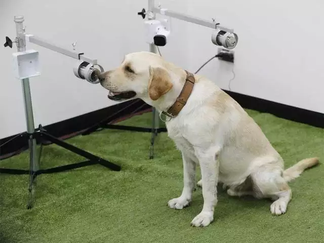
The race to improve diagnosis and treatment for AD is on. In 2023 there was even a $200,000 reward advertised for anyone who can prove that dogs can sniff out Alzheimer’s disease. Whilst MRI and PET molecular imaging of beta-amyloid and tau proteins are perhaps more promising, the cost and invasive nature of such methods precludes practical clinical applications(3).

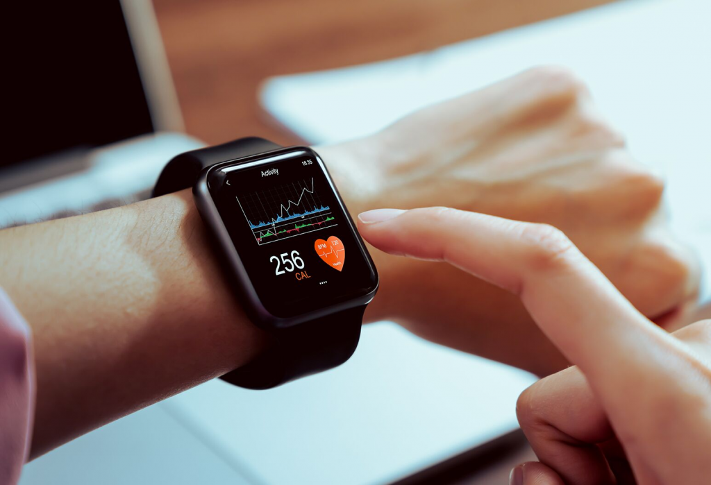
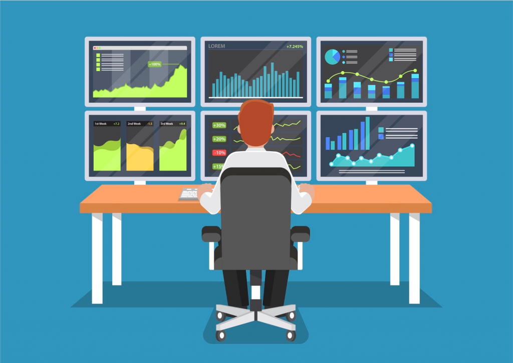
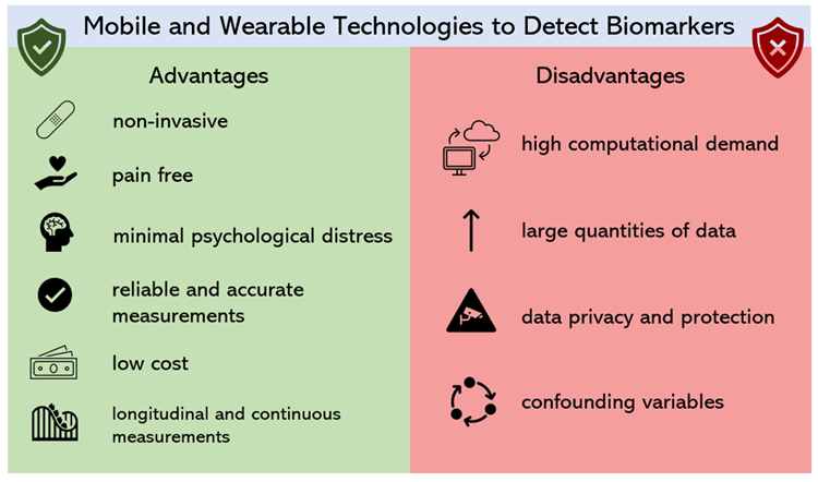
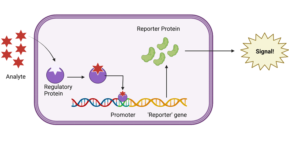
Interestingly, digital consumer technology can overcome these limitations with the added benefit of pre-symptomatic detection. What’s amazing is that cognitive, behavioural, sensory, and motor biomarkers can aid detection of AD 10 to 15 years prior to effective diagnosis(4). This allows leverage of existing technologies and sensors such as smartphone microphones and GPS systems.

Watch this quick video I put together to find out more…

Original video, inspired by Kourtis et al (2019).

Innovation or Invasion?

Whilst I do own both a smartphone and an (albeit old) smartwatch, the concept of these devices continuously monitoring everything from my speech to my walk and even the way I view an Instagram post, makes me slightly uneasy. However, I can’t deny the advantages of personal continuous monitoring for public health. The question is where do we draw the line? Could we be heading down a path where continuous passive monitoring involves cameras wired up in our home, even in our toilets?! (apparently yes! Find out more here).

I think the answer is purely personal – for some people continuous passive monitoring may be the difference between life and death, for others it might feel a little too 1984. For people experiencing cognitive decline, informed consent may complicate matters further. According to the Mental Capacity Act (2005), many people with dementia may be considered ‘Incompetent adults’ (I’m not a fan of that term) if they fail to understand the device, and cannot remember or communicate the reasoning for use. This means they would be legally unable to consent. Thankfully, the increasing nature of wearable technologies may mean that, in the future, many people will own devices decades before they are at high risk of AD, and thus can choose (at legal capacity) whether or not to install bio-monitoring software. Of course, if these devices continue to be solely commercial, then the financial accessibility of these devices may be limited – which is a whole other debate in itself.

Original image: the pros and cons of digital biomarkers.

Summary

Given the increasingly technological cultural landscape – our access to devices capable of passive monitoring is increasing. Considering the UK Alzheimer’s epidemic and our ageing population, it seems a waste to not make use of the endless potential health benefits of these devices (especially as many already monitor us for consumer metrics anyway)! Although the degree of monitoring may seem invasive, I think Alzheimer’s is a far bigger threat to our personal privacy and autonomy. Such developments could help people communicate with their families and manage symptoms before its too late.

References

  1. Dementia vs. Alzheimer’s Disease: What Is the Difference? | alz.org
  2. Kourtis, L.C. et al. 2019. Nature. 2, 9. https://doi.org/10.1038/s41746-019-0084-2
  3. Bao, W. et al. 2021. Front. Aging Neurosci. https://doi.org/10.3389/fnagi.2021.624330
  4. Vrahatis, A. G. 2023. Sensors. 23, 9. https://doi.org/10.3390/s23094184
  5. Stringer, G. et al. 2018. Int J Geratr Psychiatry. 33,7. https://doi.org/10.1002%2Fgps.4863
  6. Sun, J. et al. 2022. Sec. Brain Imaging and Stimulation. 16. https://doi.org/10.3389/fnhum.2022.972773

Navigating the Ethical Concerns Surrounding Foetal Bovine Serum

The concept for my summative blog emerged during my dissertation lab project, where I used HepG2 cells to assess the hepatoprotective impacts of chlorogenic acid against paracetamol induced liver toxicity. During this process I became familiar with cell culture, treating and assaying them most days, but when I came to seed the cells myself and prepare new culture medium, my attention was brought to foetal bovine serum. When researching the topic I found interest in the ethical debate surrounding the production of foetal bovine serum and the demand for alternatives.

Foetal bovine serum (FBS) is a key component of cell culture medium and is used extensively for research applications. It contains essential hormones and growth factors providing a rich culture system; supporting the widest range of cell types, including cell lines and primary cells.

Figure 1: European Serum Products Association Summary of FBS Production.

Although figure 1 provides a thorough overview of the FBS production process, it fails to describe the actual harvest, the point of most concern. FBS is typically obtained by the cardiac puncture of cow foetuses sourced from cows found to be pregnant after slaughter. This is carried out once the foetuses are declared deceased and without the administration of anaesthesia.

An article from 2002 covers the proposed reasons behind the ethical concerns surrounding FBS production and the potential for foetuses to experience pain during the process. One of the primary ethical concerns regarding the harvesting procedure involves the resistance of mammal foetuses to anoxia; the complete loss of oxygen supply to the body or brain. Mammals possess protective mechanisms allowing maintained brain function even after oxygen deprivation. These mechanisms include the ability to meet energy requirements via anaerobic respiration, the prioritisation of blood flow to critical organs such as the brain, and a reduced oxygen demand in the foetal brain, compared to adults. It is expected that the bovine foetuses have normal brain function at time of cardiac puncture, allowing us to assume that they are aware of the procedure and experience pain as a result. The paper then goes on to explore the timeline by which the cow foetus develops nociception and the ability to experience pain.

Since this paper is now over 20 years old I looked for more recent advancements. This led me to an article addressing the animal welfare ethics concerning FBS production and how the industry has altered it’s practises to avoid such concerns.

To summarise, the article discusses the debates regarding foetal sentience and consciousness, raising concerns about potential suffering during the procedure and proposes steps to minimise harm. They specifically make reference to the European Food Standards Agency (EFSA) recommendation to immediately open the uterus, stun and kill the foetus upon detecting pregnancy in a slaughtered cow. This ensures the foetus is unconscious and therefore doesn’t experience pain during the blood extraction. They suggest increased monitoring within the serum industry to ensure compliance with ethical standards and that by implementing such measures, the industry can demonstrate its commitment to ethical practise. Like the 2002 paper, they also conclude that it is time for researchers to adopt more modern approaches to human cell culture-based research and to move away from the use of FBS.

Figure 2: Summary of Emerging Alternatives to FBS

To conclude, at present time the production of FBS is deemed to be up to ethical standards providing individual producers follow legislation and guidelines provided. It’s clear that to fully address the concerns around animal welfare, alternatives to FBS are required. By adopting such alternatives, we can achieve greater scientific accuracy whilst demonstrating stronger ethical consideration.

References:

Jochems, C.E.A., van der Valk, J.B.F., Stafleu, F.R. and Baumans, V. (2002). The use of fetal bovine serum: ethical or scientific problem? Alternatives to laboratory animals : ATLA, [online] 30(2), pp.219–27. doi:https://doi.org/10.1177/026119290203000208.

McCann, T.J. and Treasure, C. (2022b). Addressing Animal Welfare Issues in Fetal Blood Collection for Fetal Bovine Serum Production. Alternatives to Laboratory Animals, 50(5), pp.365–368. doi:https://doi.org/10.1177/02611929221117992.

The ‘Imitation Game’: Revolutionising The Future Of Soft Robotics.

I came across a TedTalk titled ‘The artificial muscles that will power robots of the future’, which made me think – robots don’t look human at all, is there really a way to do that? An artificial muscle which is cheap, easily constructed and versatile? It cannot be?!

Many advances to improve the look of robots are already ‘in motion’. The invention of the Squidbot in 2020, was made to mimicked the swimming of a squid. The Pneumatic Artificial Muscle (PAM) was designed to aid the rehabilitation of patients diagnosed with polio. However, the HASEL actuator stood out to me, due to its versatility with its use, and the potential it has for improving both quality of life and broadening professional career paths for athletes. The concept of the HASEL (Hydraulic Amplified Self-healing ELectrostatic actuators) provides an unseen side of robotics, making the newly advancing AI robots look last season!
Image of the Peano-HASEL actuator
Their first prototype, Peano-HASEL actuator, was designed to contract linearly after electrical stimulation. The dynamic properties mimic those of an anatomical muscle. The elastomeric shell makes it easily recoverable from any force, comparative to the quick relaxation of the human muscles. The voltage applied to either end causes the liquid to migrate, producing a ‘contraction’. I believe soft robotics will go a long way for creating future real-life animatronics, which could even have the potential to function better as a result.
The Planar-HASEL being used as the bicep muscle to throw a ball.
It doesn’t stop there! The Planar HASEL actuator was made to lift heavy objects. Electrical charge is applied to the insulating liquid, causing stretching of the polymer and horizontal shrinking, making it move vertically. The future for this specific design is bright; It could completely replace upper arm prosthetics for everyday use! It can even collect enough force to throw a ball in the air, supporting my opinion: the benefits of this development will be transitional through generations!
The Quad Donut HASEL prototype can run on superspeed – put it this way, when I saw the video I couldn’t tell it was moving! It has a circular shape, and each ‘pod’ lies on top of one another. As the voltage is applied, liquid migrates to either side causing the ‘Donut’ shape. One improved function is the smoothness of motion. The mechanics allows for great control, helpful for picking up delicate objects. If this doesn’t show elegance, I don’t know what does! After seeing this photo, my thought was that if this is simple HASEL technologies, surely it can be developed for both everyday and professional use? Not only improving mental health but broadening delicate skills such as chess! I don’t see the harm in trying to improve already invented prosthetic designs! And it gets better! The use of the thermoplastic polymers for the bags provides a low-cost production alternative to current prosthetic materials such as carbon fibre which, although durable, are extremely stiff. (Rothemund et al., 2020). Of course, with every invention, there are ALWAYS drawbacks, especially one with increasing pressures to be ‘perfect’ in today’s research era. One is the lack of temperature resistance. My proposal is to add a thin heat-resistant film, to prevent it becoming rigid whilst also becoming durable. Incorporating outer metal gratings for flexibility would allow contraction and protection. This increases sustainability of the HASEL, whilst maintaining the excellence of mechanisms intact. The hope is that HASEL will lend a helping hand to those who need it!
Video showcasing examples of all the prototypes
References: Rothemund, P., Kellaris, N., Mitchell, S.K., Acome, E. and Keplinger, C. (2020). HASEL Artificial Muscles for a New Generation of Lifelike Robots—Recent Progress and Future Opportunities. Advanced Materials, 33(19), p.2003375. doi:https://doi.org/10.1002/adma.202003375.

Moral Reflections: Organ Transplants in the Shadow of Frankenstein

An abstract illustration depicting the creation of the monster Frankenstein.

Organ transplants have revived the dead so it’s not all that different to the classic gothic tale of Frankenstein by Mary Shelley. My fascination for the tale as well as the tissue engineering and stem-cell lectures led to a deep dive into this topic. The tale depicts Doctor Frankenstein’s creation of life from post-mortem organs that haunt him, but now, centuries later in modern medicine, it suggests a futuristic prophecy.


Creating Life in the Lab

Regenerative medicine is still in its early phases but there has been tremendous advancement. Pluripotent stem cells are cells that can differentiate into any type of cell in the body, so work has been conducted to grow organs from scratch with these cells. However, it’s difficult to replicate the intricate tissue organisation and complexities of organs. Just like Doctor Victor Frankenstein, scientists are itching to discover the secrets of “life and death” and learn how to “renew life”.

Brain organoids are helping scientists launch sophisticated studies of how diseases develop in the brain. (Illustration by Hindawi)

Although not applicable for transplantation, miniature organoids (‘organ-like’) have been created around various laboratories to replicate hearts, kidneys and more. Biomedical scientists such as myself can use organoids as models when testing a new drug, without harming patients. It can help understand and assess drug safety before a new drug is given to humans in clinical trials to save a lot of money and time spent in trials. In the Netherlands these organoids were used to personalise medicine for cystic fibrosis patients who were treated based on how an organoid grown from their cells behaved in response to drugs.

Organ Transplant

Transplantation of human organs is regarded as one of the key markers of 20th-century medicine. The first successful kidney transplant was performed in 1954 and since then the demand for transplant has skyrocketed. Thus, doctors have attempted to put artificial and animal-derived organs into patients. On the one hand scientific curiosity such as this has advanced human knowledge and life expectancy, but it makes me wonder at what point can curiosity go too far?

Ethics

I always wondered how medical professionals could pass judgement and determine who is worthy of a transplant. I found that in the UK strict criteria exist, where some criteria are objective (e.g. blood type) while others depend on ethical judgement. It raises questions like:

    • Should people’s lifestyle choices (smoking, obesity, drug use) that implicated their organs be given an opportunity for organ transplant?
    • Should prisoners receive a transplant?
    • Should people get a chance to get a second transplant?

David Bennett with his surgeon who transplanted a genetically modified pig heart at University of Maryland Medical Center.

David Bennett was the world’s first recipient of genetically modified pig heart after being denied a human heart. Media suggested the denial was due to his advanced heart failure while his son declared it was due to his non-compliance in managing his health. This debate continued after the surgery as news about his conviction after stabbing a man in the 1980s resurfaced. The unfortunate victim’s sister argued she wishes the modified heart “had gone to a deserving recipient.”

Final Thoughts

I think Frankenstein’s key message is that curiosity and ambition need to be met with caution and compassion, which I think has also shaped current ethical guidelines. Researching the complex interplay of science and ethics of organ transplant shifted my perspective into a more comprehensive and holistic one. 

Engineering the Invisible: the link between microbes and biosensors

As a natural sciences student, my degree is all about interdisciplinary science. I’m currently taking a third-year microbiology module, so when I found out I could write this blog on any subject related to engineering replacement body parts I was determined to find a link between my fascination with microbiology and an application to human health.

Using bacteria in biosensors

During the two lectures on sensors an ‘artificial pancreas’ was discussed, which combines a blood glucose sensor with an insulin delivering device to precisely monitor and modulate the blood glucose concentration in people with type I diabetes. This innovative technology inspired me to investigate if there could be a link between biosensors and microbiology. I quickly found an article describing bacteria biosensors. Bacterial biosensors, known as whole cell bacterial biosensors (WCBBs), work by utilising bacteria’s natural system of recognising a molecule and responding to it by producing a protein. The biosensors use genetic engineering (GE) to engineer the bacteria to recognise specific molecules and produce specific reporter proteins in response. These reporter proteins then act as a signal for the presence of the analyte which can be detected by a computer interface.

Diagram adapted from Bacterial Biosensors: The Future of Analyte Detection (asm.org), summarising the key processes in a WCBB. Created with Biorender.com.

Bacterial biosensors in practice

These WCBBs are useful in a biomedical setting, allowing the detection of molecules which may be indicative of disease, such as WCBBs which can detect cancerous DNA. However, for these biosensors to work, they must have access to the body. Engineers at MIT have developed an ingestible bacterial biosensor capsule, which they hope will soon be clinically applicable, allowing the continuous monitoring of gastrointestinal health over weeks. Therefore, through the collaboration of biological and material engineering, these MIT researchers have facilitated the use of these biosensors in the body.

The ingestible bacterial-electronic sensor from MIT school of engineering.

The debate surrounding genetic engineering

Greenpeace has a long history of protests against GE crops.

GE is often a controversial topic, with negative media coverage most commonly against genetically engineered crops. The main concern is the effect of GE species in the event of their uncontrolled release into the environment. Professor Caroline Ajo-franklin, who runs a biosciences research group at Rice University developing WCBBs, describes the need for tactics to prevent such an environmental release of GE microbes through physical containment. Overall, I believe that using proper regulation to perceive and mitigate risks, GE is a clear force for good in biomedical research. The discussion surrounding GE and the prevailing public scepticism towards it also highlighted to me the need for effective communication of research to the public, and the importance of open debate of new technologies and there applications.

The importance of seemingly unimportant links

What struck me most whilst researching this topic was the variety of bacterial use in engineering replacement body parts, with the WCBBs discussed above only a tiny snapshot of potential. I could have written about the use of bacterial biomolecules as biomedical scaffolds for human organ tissue culture, or the use of algal cells to help restore a man’s vision, or the complex role of the gut microbiome in our health. However, much like the word limit of this blog, the world of science research is limited by resource availability and funding. It’s impossible for a researcher to explore every rabbit hole, which is why I believe that interdisciplinary collaboration is integral to the future of all areas of research, and it may be the links between seemingly unrelated subjects which drive future technological breakthroughs.

Sensors in Prosthetics

Following a brief introduction in our lectures to sensors and prosthetics, I was inspired to learn more. Like many people, I had never considered all the limitations that affect prostheses users. I wanted to see how sensors could help make bionic limbs work as well as human limbs.

Controlling prostheses

In lectures we learnt about electromyogram (EMG) sensors which are used to control prostheses. To find out more I spoke to an engineering student who has produced a low budget EMG sensor as part of his year 3 project.

Short interview on EMG – Picture sourced from Electrotherapy for MSP

Improving Touch

When I think of a prosthesis, I think of a replacement body part. However, as the human body is deeply complex with many integrated systems to allow us to survive and interact with our environment, prostheses simply do not have the same functionality as human limbs. One thing I thought about was the extent to which upper limp amputees can experience human touch and their surroundings through their prosthesis.

Excitingly, by using a MiniTouch device, which can be integrated into existing prosthetic limbs, thermosensitive prostheses can be produced. Experimentally this device has been successful at allowing the user to decipher between objects with different temperatures. This technology helps prosthesis users to sense human touch akin to the feeling we get through our fingertips and increases sensation to help with motor control. However, the complete sensation of human touch still cannot be experienced by the user as human touch is a lot more complex than heat. For example, thermal sensors cannot convey to the user the texture of another person’s skin, although I was pleased to learn researchers are making progress in the development of tactile sensors.

The MiniTouch device technology has been developed further here at the University of Southampton. Researchers helped prosthetic wearers feel ‘wetness’ so both dexterity and motor control can be improved.

Improving Grip

The Southampton tube sensor

Sensors can also be utilised to improve the dexterity of prostheses users by preventing objects being dropped. This can be achieved by detecting when a slip is occurring. One option for slip detection is shown here. I found the reuse of existing technology and use of simple parts fascinating. It is made from a hearing aid microphone within a tube in contact with the grip surface. Slippage results in vibration signals which are transmitted to the air within the tube and onto the microphone. Airborne noises and interference signals can be thresholded out.

Controlling Orthoses

Orthoses are like prostheses but are designed to improve the functionality of movable parts of the body. There are many uses including rehabilitation and assisting daily activities. The mPower 1000, for example, is a neuro-robotic arm brace that fits like a sleeve on a person’s arm. It has sensors that can detect even a very faint muscle signal and the mPower 1000 provides motorised assistance in response. It is intended to increase arm movement affected by neuro-logical conditions such as stroke, spinal cord injury, multiple sclerosis, cerebral palsy, muscular dystrophy, and traumatic brain injury. Ethically, I worry if orthoses are widely used by able bodied people, it could have an untold impact on society and potentially even lead to weaponisation of the human body.

My Final Thoughts

It is really important to continue developing tactile sensors so protheses can become more like human limbs. Researching this topic has changed my perspective as I now understand we are a long way from having prosthetics which work as well as natural limbs however there is a great deal of ongoing research to improve prosthetics.

Stem Cells: The Key to Conquering Autoimmune Disease?

After Nick Evans’ lectures on stem cells and regenerative medicine I was intrigued with the concept of using stem cells to treat autoimmune disease. My mother suffers from rheumatoid arthritis and I was under the impression that such conditions had long been considered incurable. I know first-hand how debilitating autoimmune disease can be, and how the side effects of current treatments are almost as severe as suffering the disease itself, which really is heartbreaking. The idea that stem cells could regress or even cure autoimmune disease seemed almost unbelievable, so I had to investigate it further.  

AI-generated interpretation of ‘stem cells in the lab’

What are stem cells?

Stem cells are the body’s solution to self-renewal and healing: ‘blank templates’ which can divide to form many different specialised cell types. Two types of stem cell have distinct therapeutic potential for treating autoimmune disease; Mesenchymal Stem Cells (MSCs), which differentiate into connective tissue cells like bone and cartilage, and Hematopoietic Stem Cells (HSCs), from which all cell types in the blood are derived. 


Living with autoimmune disease

Autoimmune diseases occur when the immune system, the body’s defense mechanism against foreign cells, cannot tell the difference between body cells and foreign ones, attacking the body’s own cells. There are various forms of autoimmune disease, commonly type 1 diabetes, where the immune system kills insulin-producing pancreatic beta cells, or rheumatoid arthritis (RA), where connective tissues in joints are destroyed. Nearly 4% of the world’s population suffers from some kind of autoimmune disease, and in the UK has been found to affect around 1 in 10 people, – impacting the lives of millions, without hope for a permanent cure. 

Hand x-ray of advanced rheumatoid arthritis

Current treatments for autoimmune disease serve only to manage symptoms and still bear significant impact on patients’ lives. In the case of RA, common treatment includes a cocktail of anti-inflammatory drugs combined with immunosuppressants. From my mother’s experience, I can personally attest as to how severe the side effects can be – constant headaches, nausea and ceaseless illness from immunosuppression – imagine having somewhere between a cold and flu in perpetuity! In fact, around half of patients quit immunosuppressants after about a year, opting to just cope with the pain instead.  

This video helped me better appreciate the impact these diseases have on the lives of individuals and their families.
AI-generated depiction of ‘stem cell treatment in vivo’

A miracle cure?

MSCs can ‘reprogram’ the immune system to stop attacking the body’s own cells and repair damaged tissues. Moreover, HSCs can be transplanted into a patient, completely replacing their dysfunctional immune system, effectively curing them of the disease. The opportunity this presents to people who have long given up hope truly is life-changing, which made me question: “why there isn’t more public excitement about stem cell-based therapies?” 


Too good to be true?

Despite their therapeutic potential, stem cell therapies are fundamentally limited by their scarcity in the body. MSCs, for example, comprise only 0.01% – 0.001% of cells in bone marrow. They must be extracted from each patient and grown at a small scale in the lab, at a cost of around $900 per 1 million cells. This presents an ethical quandry; treatment remains prohibitively unaffordable for the majority of those affected. 

My Thoughts

The advent of stem cell therapy is an exciting prospect for treating autoimmune disease, for which I have great hopes. I have come to better appreciate the importance of understanding individuals’ experiences rather than fixating solely on science, and I hope that these considerations are made with stem cell developments to prevent their misuse.

Are pigs the answer to the organ shortage?

Organ Transplantation

In 2022-2023 it was estimated that 4600 transplants were completed thanks to thousands of donors in the UK. However, it is also estimated that 7000 people were on the waiting list, almost 1.5x the number of transplants completed. Tragically, from this, 430 died waiting for an organ last year. This is a huge issue in modern medicine that needs to be tackled immediately. The NHS are advertising the importance of being an organ donor, in May 2022 the UK introduced an opt-out system. Each country in the UK has similar legal implications surrounding this subject; which states if you are over 18 and have not opted-out and aren’t in an excluded group, you are deemed authorised for organ donation. Scotland differs a little bit whereby you are deemed authorised at 16 and assume consent if not opted-out. This has increased the donor rate by 50% in 5 years and is hoping to double in 10.

When Death Turns Into Life

Keeth Reemsta

I was first interested in this subject after reading Merzrich’s When Death Turns Into Life, which discusses dilemmas of transplant surgeons as well as their breakthroughs. Reading the trials regarding xenotransplantation pricked my interest and prompted me to research further. The efforts of Keith Reemsta, a risk-taker determined to transplant primate organs into humans was particularly fascinating. One case was that of 43-year-old Jeffery Davis, who was in heart failure and end stage renal disease and could not live on short-term dialysis. Reemsta transplanted both kidneys still connected to the vena cava and aorta and treated with the available immunosuppression medication available in 1963. Davis sadly died a month and a half later from pneumonia and not rejection. Reemsta continued transplanting a further 13 patients who enjoyed 9-60 days of extended life. I respect how driven Reemsta was to move forward his research, though in 1965 this halted when chronic dialysis became available.

Primates vs Pigs?

There is evidence now that attempting to transplant chimpanzee organs is unsustainable as they are endangered, difficult to breed, have one offspring at a time, expensive to care for and exposes humans to xenoviruses due to the homology in genetics between the species. There is also an ethical debate regarding whether these animals are too much like humans, which we would raise purely to harvest organs from, where do we draw the line?

Alternatively, pigs could be used, which have more reason to be use instead, such as easy breeding, big litters, appropriate size, fair genetic homology, cheap and more socially acceptable. This is in the sense that pigs are already harvested for pork products, why could we not harvest their organs as well to help the global shortage of organs. I think this would be a major breakthrough in research. However, using pigs could cause debates from a religious standpoint, in Islamism this would be seen as a betrayal of their religion. One of the biggest issues with using pigs is the presence of alpha-gal epitope, a protein non-primate mammals possess which could lead to rejection. Since the discovery of CRISPR/Cas9, George Church has successfully generated pigs with inactivated PERV elements, causing the threat of xenoviruses to be diminished, but is still yet to get FDA-approved.

In the book there was a lovely phrase, which I believe sums up the research in xenotransplantation perfectly:

‘Xenotransplantation is just around the corner, but it may be a very long corner’ – Sir Roy Calne 1995

Below is an interesting video about why pigs should be used for future organ donation, exploring how we may be able to genetically-modify them to prevent rejection through Chimeras.

Acknowledgements

Mezrich, Joshua D. When Death Becomes Life : Notes from a Transplant Surgeon. New York, Ny, Harper, An Imprint Of Harpercollinspublishers, 2019.

NHS. “Organ Donation and Transplantation.” NHS Blood and Transplant, 2022, www.nhsbt.nhs.uk/what-we-do/transplantation-services/organ-donation-and-transplantation/.

“Organ Donation Laws.” NHS Organ Donation, 2016, www.organdonation.nhs.uk/helping-you-to-decide/organ-donation-laws/.

“Improved System of Organ Use to Save Lives.” GOV.UK, www.gov.uk/government/news/improved-system-of-organ-use-to-save-lives#:~:text=The%20opt%2Dout%20change%20to.

Mohd Zailani, Muhammad Faiq, et al. “Human–Pig Chimeric Organ in Organ Transplantation from Islamic Bioethics Perspectives.” Asian Bioethics Review, 16 Nov. 2022, https://doi.org/10.1007/s41649-022-00233-2.

A reflective journey into the profound history and motivations behind anatomical studies and it’s role in the future medical curriculum.

The corridors of the Education wing of the hospital were long and dimly lit, as though the very building bore the solemnity and calm monotony of its workers inside. Apron on and into the lab I went, greeted by the sharp air which washed the senses and clung to clothes; formaldehyde would be the odour to my education for that day. I explored specimens laid out in-front of me through gloved hands, my nerves dissipating in the pursuit of knowledge and yet a silent edge to the room remained, the taboo feeling still choking speech.

My first time stepping into an anatomy lab was an experience like no other. It’s a moment filled with a complex blend of emotions; curiosity, apprehension, and a profound sense of respect for the journey upon which you’re about to embark. Reflecting on this; the presence of the surgeon, his demeanour, his depth of experience and the way he handled the specimens with such delicate precision put me at a surprising sense of ease. It allowed me to see past extraneous detail and appreciate the beauty in the anatomy and the implants used to restore vital function. This kinaesthetic experience cemented the knowledge I’d gained over the course of my academic career. I wondered to myself, how did the history of anatomy lead up to this moment today, and what will it be like for future students such as myself?

A Historical Perspective

The scientific motivations behind anatomy labs and the pursuit of medical knowledge are strongly intertwined with the ethical considerations that have evolved over time. Cadaveric dissection has roots in 1600BC Egypt (tracing back to the Edwin Smith Surgical Papyrus) and Ancient Greco-Roman philosophers such as Hippocrates and Galen. However, it was during the Medical Renaissance (circa 1400-1700CE) that a pivotal shift towards empirical research and direct observation began, with prolific figures such as Andreas Vesalius challenging misconceptions and laying the foundations for modern anatomy. This period also incited ethical debates on the use and procurement of cadavers, which had previously been sourced from wherever available (legal or otherwise) leading to legislation such as the Murder Act 1751 stipulating that only corpses of executed convicts could be used for dissection, followed by the Anatomy Act 1832 regulating institutional licenses to practise anatomy.

In the modern era, anatomical laboratories serve as essential educational tools for aspiring doctors and are tightly regulated (Human Tissue Act 2004), a lesson learned from history intended to uphold respectful practices and promote ethical research (sometimes remaining an issue). These considerations are now at the forefront of study ensuring respect for donors and strict adherence to consent protocols.

The future of medical studies

There exists conflict surrounding the future of anatomical study, and whether it involves the potentially-archaic use of cadavers at all. As a result of modern technology, devices such as large interactive screens or virtual reality (VR) provide a low-cost alternative to expensive laboratories, reducing the expense of cadavers themselves, the equipment to store and maintain them, the toxic fumes from formaldehyde, and permit repeatability of dissection procedures without cost or irreparable damage to the specimen – simply press reset.

From my experience, this would dehumanise and detract from the solemnity and respect of learning practical anatomy, reducing exposure for the benefit of fiscal expenditure but at the cost of experience – which is quintessential for any practicing doctor. But what does an aspiring doctor think?

If I could leave one last quote in the minds of those interested in this topic, it would be from Paul Kalanithi, the author of When Breath Becomes Air.

Pioneering the Pulse: The Future of Artificial Hearts

In the intricate landscape of medical science, perhaps no feat is as remarkable as the creation of artificial hearts. These remarkable devices stand as a testament to human ingenuity and the relentless pursuit of innovation in healthcare. As we stand at the cusp of a new era in medicine, it’s important to explore the history, current challenges, and the promising future of artificial hearts.

A Journey Through Time: The History of Artificial Hearts

The genesis of artificial hearts can be traced back to the 1950s when Dr. Paul M. Zoll developed the first external pacemaker. This monumental achievement laid the foundation for further advancements in cardiac care. However, it wasn’t until 1982 that Dr. Robert Jarvik’s creation, the Jarvik-7, became the first artificial heart implanted in a human. Though it was initially intended as a temporary measure, it marked a significant milestone in medical history.

Jarvik-7: the first artifial heart implanted in a human

The Present Landscape: Temporary Solutions Amidst Growing Challenges

Today, artificial hearts primarily serve as a bridge for patients awaiting heart transplants. However, the demand for heart transplants far exceeds the available supply. One of the most significant challenges facing artificial hearts is the interaction between platelets and artificial surfaces, which triggers the activation of contact proteins and leads to coagulation. Consequently, patients require therapeutic intervention, often in the form of medications, to mitigate these effects. However, despite these efforts, there are inherent limitations on the duration for which artificial hearts can be utilized due to this phenomenon.

In the UK, the British Heart Foundation reports that over 900,000 people live with heart failure, with an estimated 1,000 new cases diagnosed each month. Each year, around 200 people in the UK are added to the heart transplant waiting list. Regrettably, due to the scarcity of donor organs, many patients face the grim reality of heart disease, facing long stays in hospital with some tragically passing away while awaiting a life-saving transplant.

Evie: admitted to hospital over a year ago and still awaiting a heart transplant

Looking Forward: The Promise of Future Artificial Hearts

As we venture into the future of artificial hearts, a myriad of technological advancements offer hope for overcoming current limitations and revolutionizing cardiac care. One area of significant progress lies in battery technology. Traditional power sources for artificial hearts, such as external batteries or power cords, present challenges in terms of mobility and infection risk. However, the development of smaller, more efficient batteries promises greater freedom and convenience for patients, allowing them to lead more active lives without constant tethering to external power sources.

Biomaterials also play a pivotal role in the advancement of artificial hearts. Researchers are exploring innovative materials that closely mimic the properties of natural heart tissue, reducing the risk of immune rejection and clot formation. These biocompatible materials not only enhance the longevity of artificial hearts but also promote better integration with the surrounding tissues, minimizing the need for anticoagulant therapy and reducing the risk of complications.

Incorporating advanced sensors into artificial hearts enables real-time monitoring of vital parameters such as blood flow, pressure, and heart rate. This continuous stream of data allows for early detection of potential issues, enabling timely intervention and improving patient outcomes.