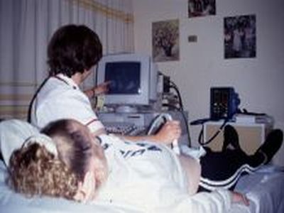
Women who had taken part in the Survey and then became pregnant were offered extra scans in the Southampton Women’s Survey ultrasound unit at Princess Anne Hospital, to follow both their progress and that of their babies.
Recruitment of pregnancies ended in 2007.

At our scanning unit we carried out an early scan at around 11 weeks to confirm the mother’s dates and check that all was well for this stage of pregnancy. We then carried out the routine 19 week scan to measure the growth of the baby and its placenta, and to check that the baby was developing normally. At 34 weeks, we measured the baby’s size and the blood flow through the cord and some of its organs including the head and kidneys, and at 36 weeks for a sample of women we measured the blood flow from the cord through baby’s liver.
Many of the women were interviewed following the 11 week and 34 week scans, during which we asked about similar factors to the initial interview, as well as some extra questions about the pregnancy; blood samples were also collected.
During the 19 week visit, many mothers- and fathers-to-be had their grip strength measured. We also collected blood and mouthwash samples from the fathers, and requested details of the woman’s parents so that we could obtain mouthwash samples from them too if possible.
Pregnancy Data Collected
| State of Pregnancy | Type of Information | Number |
|---|---|---|
| Early pregnancy (11 weeks’ gestation) | Questionnaire | 2,867 |
| Heel scan | 1,897 | |
| Ultrasound scan of baby | 2,517 | |
| Mother’s blood samples | 1,985 | |
| Mid pregnancy (19 weeks’ gestation) | Grip strength | 1,499 |
| Father’s grip strength | 1,401 | |
| Father’s mouthwash sample | 1,655 | |
| Mother’s parents’ mouthwash samples | 1,407 | |
| Ultrasound scan of baby | 3,033 | |
| 3D ultrasound scan of baby’s femurs | 517 | |
| Late pregnancy (34 weeks’ gestation) | Questionnaire | 2,647 |
| Heel scan | 2,141 | |
| Ultrasound scan of baby | 3,050 | |
| 3D ultrasound scan of baby’s femur | 490 | |
| Mothers’ blood samples | 2,309 |
As part of our studies of the baby’s bone development (detailed below), many mothers were invited to have a scan of their own heel bone during their visits, and to have a 3-D ultrasound scan of their babies’ femoral bone size at 19 and 34 weeks. This information has been linked with the mother’s intake of calcium and vitamins to assess how much of the calcium in a baby’s bones comes from the mother bones and how much from the food that she eats.
