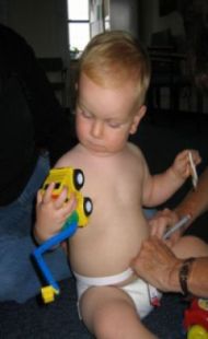
Our team at Princess Anne Hospital were kept very busy with the arrival of the SWS babies. Shortly after birth, usually at the hospital, our SWS research nurses measured the newborns. The children were then followed-up with home visits at the ages of 6 months, 1 year, 2 years and 3 years to assess their growth, diet and allergies. We also asked about the children’s illnesses, sleep and activity, and many other factors.
4 Years
Over 1,000 children came into our clinic at Southampton General Hospital for a scan to assess their bone development. We also assessed their growth and grip strength and collected data on their diet, illnesses and levels of physical activity. Some children also had home visits to measure their thinking skills.
With the mother’s consent, we also liaise with the Central Health Clinic in Southampton to record height and weight measurement data collected in all local schools when the children are four-five years old.
6-7 Years
Over 2,000 children were visited at home by our research nurses. They collected information on diet, respiratory health and thinking skills. More than 1,000 of these children subsequently visited the Southampton General Hospital for a bone scan (DXA) and many received detailed assessment of their lung function and grip strength measurements. Our assessments made at this age are combined with the findings from earlier in the children’s lives to give us a detailed picture of the factors that increase the risk of asthma and other lung problems, and how thinking skills develop.
8-9 Years
Children were invited to attend a clinic at the Princess Anne Hospital for a detailed assessment of their cardiovascular structure and function as well as a bone scan (DXA) and grip strength measurements. More than 1,000 children were seen at these clinics.
11-13 Years
We took photographs of the children’s teeth to assess tooth enamel. They had bone scans, thinking skills and grip strength assessments. An exercise step test assessed physical fitness, and a cardiovascular assessment. Some children kindly provided blood samples at this visit.
Bone scanning and osteoporosis
Building on the sub-study of 1000 babies who were scanned shortly after birth (see Additional Studies), over 1000 SWS children were invited to have a DXA scan at four years old, at 6-7 years old and 8-9 years. We scanned the children in the 11-13 year follow-up. The children were measured and weighed, and data collected about the child’s diet, activity and health. The information gained from this research helps us identify ways to improve bone health in children and thus reduce the number of fractures caused by osteoporosis in later life. Findings from our bone studies have led us to develop the MAVIDOS study
Allergy testing
We tested for allergies in our SWS mothers and children at the 1 year follow-up visit; and then tested the children again at 3 years and 6-7 years. We look to see if they are sensitive to dog, cat, egg, milk, grass or house dust mite.
We have found that whether the parents suffer from asthma or eczema influences the child’s risk of these illnesses, and that generally if the mother smokes the child is at greater risk of various respiratory problems.
Physical activity measures
Additionally, at 4 and 6-7 years, many children’s activity levels were measured by an Actiheart monitor. Over 500 child and mother pairs have worn this for up to seven days in the week following their 4 year visit, and nearly 700 children following their 6-7 year visit, with some mothers wearing them as well. This helps us to understand how physical activity affects the body composition of the children, and also how the mother’s physical activity levels, and other factors, influence her child’s physical activity.
Cardiovascular structure and function
At the 8-9 year visit, we conducted a detailed cardiovascular assessment of the children. This included a variety of measures including intima media thickness, flow mediated dilatation and pulse wave velocity as well as more common measures such as blood pressure. Some children also received an echocardiography assessment. All these measurements enable us to see how the children’s cardiovascular system is developing and to find out what factors earlier in their lives, even before birth, influence that development. A sub-sample of 250 children received a cardiac MRI scan which allowed us to look at the heart in great detail. At 11-13 years we conducted an exercise step test and took pulse wave velocity measures.
